

Summary: Hearing their mother’s voice may help premature babies develop stronger language pathways in the brain. In a groundbreaking study, preemies who regularly listened to recordings of their mothers reading had more mature language-related brain connections than those who did not.
MRI scans revealed significant improvement in the left arcuate fasciculus, a white-matter tract essential for language processing. The findings suggest that maternal speech exposure, even through recordings, could meaningfully enhance early brain development in preterm infants.
Key Facts:
- Language Pathway Growth: Babies exposed to recordings of their mothers’ voices showed more advanced development in a key brain tract for language.
- Early Intervention: Just a few weeks of nightly recordings produced measurable neurological differences before hospital discharge.
- Practical Impact: The approach offers a simple, low-cost way to support cognitive and language development in hospitalized preemies.
Source: Stanford
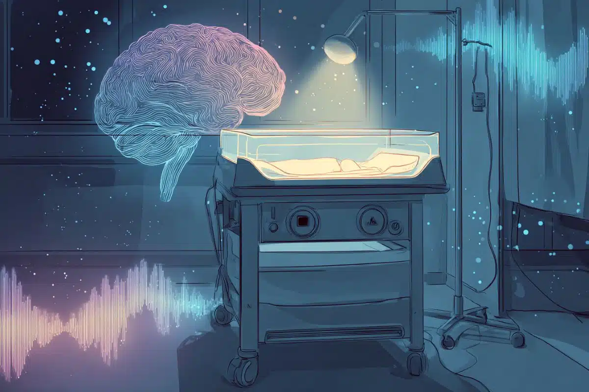
Hearing the sound of their mother’s voice promotes development of language pathways in a premature baby’s brain, according to a new Stanford Medicine-led study.
During the study, which is publishing online Oct. 13 in Frontiers in Human Neuroscience, hospitalized preemies regularly heard recordings of their mothers reading to them.
At the end of the study, MRI brain scans showed that a key language pathway was more mature than that of preemies in a control group who did not hear the recordings. It is the first randomized controlled trial of such an intervention in early development.
“This is the first causal evidence that a speech experience is contributing to brain development at this very young age,” said the lead author, Katherine Travis, PhD, who was an assistant professor at Stanford Medicine when the study was conducted and is now an assistant professor at Weill Cornell Medical School and Burke Neurological Institute.
“This is a potentially transformative way of thinking about how to approach neonatal care for promoting better language outcomes in children born prematurely.”
The study’s senior author is Heidi Feldman, MD, PhD, the Ballinger-Swindells Endowed Professor in Developmental and Behavioral Pediatrics.
Premature babies — born at least three weeks early — often spend weeks or months in the hospital, typically going home around their original due dates. During hospitalization, they hear less maternal speech than if they had continued to develop in utero.
Parents can’t usually stay at the hospital around the clock; they may have older children to care for or jobs they must return to, for example. Preemies are at risk for language delays, and scientists have suspected that reduced early-life exposure to the sounds of speech contributes to the problem.
The researchers decided to boost preemies’ exposure to their mom’s voices during hospitalization. They did this by playing recordings of the mothers speaking, a total of 2 hours and 40 minutes a day, for a few weeks at the end of the babies’ hospital stays.
“Babies were exposed to this intervention for a relatively short time,” said study co-author Melissa Scala, MD, a clinical professor of pediatrics and a neonatologist at Lucile Packard Children’s Hospital Stanford. “In spite of that, we were seeing very measurable differences in their language tracts. It’s powerful that something fairly small seems to make a big difference.”
Hearing develops in utero
Fetal hearing begins to develop a little more than halfway through pregnancy, around 24 weeks into what is normally a 40-week gestation period. As the fetus grows, the uterus expands and the uterine wall thins.
Late in pregnancy, more sounds, including the mother’s conversations, reach the fetus. At birth, full-term newborns recognize their mother’s voice and prefer the sounds of their parents’ native language to other languages, prior research has shown.
These factors suggest that listening to Mom’s voice contributes to brain maturation in the latter half of a full-term pregnancy. “The body isn’t going to waste energy developing hearing so early if it’s not doing something important to program the brain,” Scala said.
Travis, Scala and their colleagues realized that by supplementing the sounds that premature babies hear in the hospital so they resemble what they would have heard in the womb, they had a unique opportunity to possibly improve brain development at this stage of life.
The 46 babies in the study were born very prematurely, arriving more than eight weeks early. Babies could join the study when they were medically stable and had “graduated” from the neonatal intensive care unit, where the sickest newborns receive care, to the intermediate care nursery, where they stay until they are ready to go home.
The babies in the study did not have congenital anomalies and had not experienced major complications of preterm birth.
The researchers recorded mothers reading a chapter of Paddington Bear, a children’s book that has been translated into many languages. Each mother made a recording for her baby in her native language.
The babies were randomly assigned to the treatment group (who heard their moms’ voice recordings) or the control group (who did not). For babies in the treatment group, the recordings were played during the night in 10-minute periods, for a total of 160 minutes each night.
By playing the recordings at night, the researchers prevented the parents from knowing which group their babies were in, ensuring that the parents’ behavior wouldn’t affect the results.
The recordings did not appear to interfere with babies’ sleep, the researchers said, noting that fetuses at the same stage of development often sleep while their mothers are awake and talking.
The babies received MRI scans of their brains as part of the usual health checks given before hospital discharge. The scans included imaging of the arcuate fasciculus tracts on both sides of the brain, which contain large bundles of nerve fibers that help process and understand sound. The left arcuate fasciculus is specialized for language processing.
Changes in a key language pathway
On brain scans, the researchers saw a significant difference in the white matter in the left arcuate fasciculus: This language-processing pathway was more mature in babies in the treatment group than those in the control group.
The right arcuate fasciculus was less affected by the treatment, which is consistent with known differences in how the two hemispheres of the brain process speech, the scientists said.
“I was surprised by how strong the effect was,” Travis said. “That we can detect differences in brain development this early suggests what we’re doing in the hospital matters. Speech exposure matters for brain development.”
The researchers are planning new studies to test whether the benefits extend to babies with medical complications, and they are exploring in more detail how the voice recordings exert their effects.
Scala also leads a team at Packard Children’s that is creating customized plans for patients so that the sounds that preemies hear in the hospital’s NICU are most conducive to promoting brain development.
Encouragement for parents
Parents of preemies often experience stress during their baby’s hospitalization, including feelings of helplessness or frustration that they cannot spend as much time with their babies as they want to.
“We’ll always support parents visiting and talking to their babies in person as much as they can,” Scala said, noting that in-person visits also offer opportunities for parents to hold their babies skin to skin, which confers its own neurodevelopmental benefits.
She also hopes parents will feel encouraged to learn that voice recordings can supplement in-person visits.
“This is a way that — even if they can’t be there as much as they want to — the baby is still hearing them and still knows that they’re there,” she said. “And the parents are still contributing to the baby’s brain development.”
Funding: The research was supported by the Eunice Kennedy Shriver National Institute of Child Health and Human Development (grants 5R00-HD84749 and 2R01-HD069150).
Key Questions Answered:
A: Premature babies often miss out on crucial sound exposure in the womb; hearing their mother’s voice may help bridge that developmental gap.
A: Researchers played recordings of mothers reading aloud to their preterm babies for about 160 minutes each night during hospitalization.
A: The scans showed that babies who heard their mothers’ voices had more mature language-related white matter on the brain’s left side.
About this language development research news
Author: Erin Digitale
Source: Stanford
Contact: Erin Digitale – Stanford
Image: The image is credited to Neuroscience News
Original Research: Open access.
“Listening to Mom in the Neonatal Intensive Care Unit: A randomized trial of increased maternal speech exposure on white matter connectivity in infants born preterm” by Katherine Travis et al. Frontiers in Human Neuroscience
Abstract
Listening to Mom in the Neonatal Intensive Care Unit: A randomized trial of increased maternal speech exposure on white matter connectivity in infants born preterm
Objective:
Early speech experiences are presumed to contribute to the development of brain structures involved in processing speech. Previous research has been limited to correlational studies. Here, we conducted a randomized trial with neonates born preterm to determine whether increased exposure to maternal speech during NICU hospitalization is causally linked to structural white matter maturation.
Study design:
We enrolled 46 neonates born preterm (24–31 weeks gestational age). Participants were randomly assigned to receive increased (T: n = 21) or routine (C: n = 25) exposure to mother’s speech. The T-group heard 10-min audio recordings of their mothers reading a children’s story two times/hour between 10pm and 6am, increasing speech exposure by 2.67 h/day. The C-group did not hear recorded speech. At near-term-equivalent age, we obtained two high-angular resolution diffusion MRI (scan 1: b = 700, scan 2: b = 1500) and T1 relaxometry scans.
We assessed mean diffusivity (MD), pre-registered primary outcome (NCT02847689), of the left and right arcuate fasciculus, tracts implicated in language processing. Secondary outcomes included fractional anisotropy (FA) and R1 (1/T1). We hypothesized that neonates randomized to the T-group would show evidence for increased maturation within the arcuate, indexed as decreased MD and increased FA and R1, compared to neonates in the C-group.
Results:
Groups were equivalent on medical and demographic variables. Linear mixed models demonstrated that compared to the C-group, the T-group demonstrated significantly lower MD in the left (scan 1: β = −0.11, Marginal R2 = 0.27; scan 2: β = −0.12, Marginal R2 = 0.33) but not right arcuate (scan 1: β = −0.06, Marginal R2 = 0.09; scan 2: β = −0.03, Marginal R2 = 0.01). The T-group also demonstrated significantly higher FA (scan 1 β = 0.02, Marginal R2 = 0.20; scan 2: β = 0.03, Marginal R2 = 0.31) and R1 (β = 0.02, Marginal R2 = 0.39) in the left but not right arcuate.
Conclusion:
Preterm neonates with increased maternal speech exposure showed more mature left arcuate microstructure, supporting a causal role of exposure to speech in brain development. Enhancing speech exposure in the NICU may benefit preterm children’s language outcomes.


