

Summary: Hippocampal neurons involved in Pavlovian learning shift their behavior and become more synchronized when a memory is being formed. The findings shed new light on the neurobiological mechanisms of memory and learning.
Source: University of New Hampshire
The phrase “Pavlov’s dogs” has long evoked images of bells, food and salivating dogs. Even though this tried-and-true model of repetitive patterns mimics a variety of learning processes, what happens on a cellular level in the brain isn’t clear. Researchers at the University of New Hampshire took a closer look at the hippocampus, the part of the brain critical for long-term memory formation, and found that the neurons involved in so-called Pavlovian learning shift their behavior during the process and become more synchronized when a memory is being formed – a finding that helps better understand memory mechanisms and provides clues for the development of future therapies for memory-related diseases like dementia, autism and post-traumatic stress disorder (PTSD).
“There are tens of millions of neurons in the hippocampus but only a small fraction of them are involved in this learning process” said Xuanmao (Mao) Chen, assistant professor of neurobiology. “Before engaging in Pavlovian conditioning, these neurons are highly active, almost chaotic, without much coordination with each other, but during memory formation they change their pattern from random to synchronized, likely forging new connecting circuits in the brain to bridge two unrelated events.
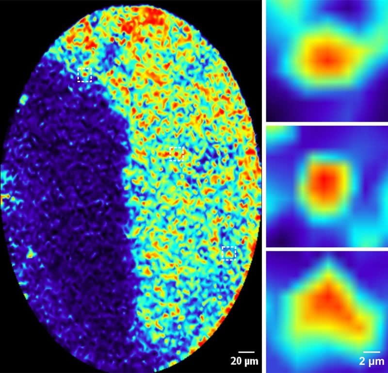
In the study, recently published in The FASEB Journal, researchers looked at Pavlovian learning patterns, or respondent conditioning, in mice. In the beginning, before any repetitive learning exercises, the mice did not know what to expect and using special imaging with an endomicroscope the researchers saw that the neural activity was disorderly. But after repeating different tasks associated with a conditional stimulus, like a tone or bell, the mice began to recognize the pattern and the highly active neurons became more synchronized. The researchers hypothesize that without forming synchronization, animals cannot form or retrieve this type of memory.
In the 1890’s, Russian psychologist, Ivan Pavlov discovered classical conditioning through repetitive patterns of bell ringing which signaled to his dogs that food was on its way and stimulated salivation. This same learned behavior is important for episodic knowledge which is the basis for such things as learning vocabulary, textbook knowledge, and memorizing account passwords. Abnormal learning processing and memory formation are associated with a number of diseases like dementia, autism, and PTSD. People who struggle with these cognitive dysfunction-related disorders may have trouble retaining memories or can even form too strong a memory, as with PTSD patients. The UNH researchers believe that understanding the fundamentals of how classical conditioning shape neural connections in the brain could speed up the development of treatments for these disorders in the future.
Contributing to these findings are Yuxin Zhou, doctoral candidate; Liyan Qiu, research scientist; both at UNH, and Haiying Wang, assistant professor at the University of Connecticut.
Funding: This work was supported by the National Institutes of Health (NIH) and the Cole Neuroscience and Behavioral Faculty Research Awards.
Source:
University of New Hampshire
Media Contacts:
Robbin Ray – University of New Hampshire
Image Source:
The image is credited to UNH.
Original Research: Open access
“Induction of activity synchronization among primed hippocampal neurons out of random dynamics is key for trace memory formation and retrieval”. Yuxin Zhou, Liyan Qiu, Haiying Wang, Xuanmao Chen.
FASEB Journal doi:10.1096/fj.201902274R.
Abstract
Induction of activity synchronization among primed hippocampal neurons out of random dynamics is key for trace memory formation and retrieval
Memory is thought to be encoded by sparsely distributed neuronal ensembles in memory‐related regions. However, it is unclear how memory‐eligible neurons react during learning to encode trace fear memory and how they retrieve a memory. We implemented a fiber‐optic confocal fluorescence endomicroscope to directly visualize calcium dynamics of hippocampal CA1 neurons in freely behaving mice subjected to trace fear conditioning. Here we report that the overall activity levels of CA1 neurons showed a right‐skewed lognormal distribution, with a small portion of highly active neurons (termed Primed Neurons) filling the long‐tail. Repetitive training induced Primed Neurons to shift from random activity to well‐tuned synchronization. The emergence of activity synchronization coincided with the appearance of mouse freezing behaviors. In recall, a partial synchronization among the same subset of Primed Neurons was induced from random dynamics, which also coincided with mouse freezing behaviors. Additionally, training‐induced synchronization facilitated robust calcium entry into Primed Neurons. In contrast, most CA1 neurons did not respond to tone and foot shock throughout the training and recall cycles. In conclusion, Primed Neurons are preferably recruited to encode trace fear memory and induction of activity synchronization among Primed Neurons out of random dynamics is critical for trace memory formation and retrieval.


