Summary: Study sheds light on how macrophages use a specific cell signalling pathway to control peripheral nerve repair following damage.
Source: University of Plymouth.
New research led by the University of Plymouth has shed light on the science behind peripheral nerve repair, by highlighting the novel function of a cell called a macrophage.
Macrophages are part of our immune system and are found in most tissues, where they patrol for potential harmful organisms and destroy them. They also play an important role in tissue repair and wound healing following traumatic injury.
The research, published today (5 February) in the journal Cell Reports, shows for the first time how the macrophage uses a specific cell signalling pathway to control the processes of nerve repair following damage. By understanding how it works, scientists hope to develop novel therapeutic approaches to enhance the efficiency of peripheral nerve repair.
Peripheral nerves connect the brain and spinal cord (central nervous system) to the rest of the human body. Damage to these nerves can cause a permanent loss of sensation in the affected area or an inability to control our muscles and move; things we take for granted in our everyday lives.
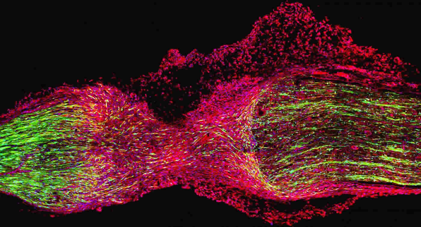
The nerves that connect our bodies to the spinal cord and brain consist of cable-like bundles of nerve projections (known as axons) that are insulated and protected by specialised cells called Schwann cells. When a nerve is injured, the two ends of the severed nerve may separate and new tissue, known as a nerve bridge, forms between the two nerve stumps.
This new tissue consists of several cell types, including migrating Schwann cells and macrophages. It is the interaction between Schwann cells and macrophages, and how this process allows the nerve projections to successfully regrow, that is the subject of the new study. This process is vital if the new nerve projections are to reach the skin and muscle, allowing for regeneration and a full recovery of nerve function.
The study, conducted in mice, shows how macrophages form a collar around the outside of the nerve bridge and secrete high levels of a repulsive nerve guidance cue called Slit3. Meanwhile, the researchers also discovered that Schwann cells inside the nerve bridge express the Slit3 interacting protein called Robo1.
Through the interaction between Slit3 and Robo1, the macrophages keep migrating Schwann cells and the regrowing nerve projections inside the bridge area. This is a vital part of the repair process; without it, the nerve projections cannot correctly get to their targets and will never reconnect with skin and muscle, leaving patients with a permanent loss of nerve function.
Lead author Dr Xin-Peng Dun, from the Institute of Translational and Stratified Medicine (ITSMed) at the University of Plymouth explains: “Peripheral nerve injuries represent a huge clinical challenge in healthcare. More than one million people worldwide suffer from peripheral nerve damage each year – and our work has exciting potential to help patients to improve the outcome of such injuries.
“We want to understand the Slit3-Robo1 signalling and utilize it to prepare engineered nerve tissue to boost peripheral nerve repair. So it was really interesting to see the key and novel role of the macrophage, and how its production of Slit3 controls cell migration and the axons’ ‘pathfinding’ in the peripheral nerve bridge.
“We know peripheral axons have the ability to grow back into their original targets after being severed, but they do not do this quickly enough when they face the problem of long nerve gaps following injury. In this study in mice, the nerve bridges being formed were around 2mm long. But in human patients the gap can be up to 5cm to fill, so it’s important for us to keep working to explore the details of long nerve gap repair and improve the clinical management of such injuries.”
Source: Amy King – University of Plymouth
Publisher: Organized by NeuroscienceNews.com.
Image Source: NeuroscienceNews.com image is credited to University of Plymouthy.
Original Research: Open access research for “Macrophage-Derived Slit3 Controls Cell Migration and Axon Pathfinding in the Peripheral Nerve Bridge” by Xin-peng Dun, Lauren Carr, Patricia K. Woodley, Riordan W. Barry, Louisa K. Drake, Thomas Mindos, Sheridan L. Roberts, Alison C. Lloyd, and David B. Parkinson in Cell Reports. Published February 5 2019.
doi:10.1016/j.celrep.2018.12.081
[cbtabs][cbtab title=”MLA”]University of Plymouth”The Key Function of Specialized Cells in Peripheral Nerve Repair.” NeuroscienceNews. NeuroscienceNews, 6 February 2019.
<https://neurosciencenews.com/macrophage-peripheral-nerve-repair-10704/>.[/cbtab][cbtab title=”APA”]University of Plymouth(2019, February 6). The Key Function of Specialized Cells in Peripheral Nerve Repair. NeuroscienceNews. Retrieved February 6, 2019 from https://neurosciencenews.com/macrophage-peripheral-nerve-repair-10704/[/cbtab][cbtab title=”Chicago”]University of Plymouth”The Key Function of Specialized Cells in Peripheral Nerve Repair.” https://neurosciencenews.com/macrophage-peripheral-nerve-repair-10704/ (accessed February 6, 2019).[/cbtab][/cbtabs]
Abstract
Macrophage-Derived Slit3 Controls Cell Migration and Axon Pathfinding in the Peripheral Nerve Bridge
Slit-Robo signaling has been characterized as a repulsive signal for precise axon pathfinding and cell migration during embryonic development. Here, we describe a role for Sox2 in the regulation of Robo1 in Schwann cells and for Slit3-Robo1 signaling in controlling axon guidance within the newly formed nerve bridge following peripheral nerve transection injury. In particular, we show that macrophages form the outermost layer of the nerve bridge and secrete high levels of Slit3, while migratory Schwann cells and fibroblasts inside the nerve bridge express the Robo1 receptor. In line with this pattern of Slit3 and Robo1 expression, we observed multiple axon regeneration and cell migration defects in the nerve bridge of Sox2-, Slit3-, and Robo1-mutant mice. Our findings have revealed important functions for macrophages in the peripheral nervous system, utilizing Slit3-Robo1 signaling to control correct peripheral nerve bridge formation and precise axon targeting to the distal nerve stump following injury.