Summary: Researchers discovered how inflammation in the womb can disrupt the maturation of nerve cells and impede an unborn baby’s brain development.
The study reveals that excessive inflammation can lead to reduced neuronal electrical activity, possibly resulting in conditions like cerebral palsy or developmental issues affecting growth, movement, vision, and hearing. Additionally, this research demonstrates that these impairments in nerve cell maturation can be detected through electroencephalography.
The team emphasizes the importance of maintaining a balanced environment for optimal brain development in fetuses.
Key Facts:
- Excessive inflammation in the womb can hinder the maturation of nerve cells, leading to reduced electrical activity and affecting a baby’s brain development.
- This condition can potentially lead to a wide range of developmental issues in babies, especially those born preterm, including cerebral palsy, emotional problems, language and cognitive delays, and more.
- The impairments in nerve cell maturation caused by inflammation can be detected using electroencephalography, a widely used clinical tool.
Source: Hudson Institute of Medical Research
It has long been recognized that too much inflammation in the womb can harm an unborn baby’s brain development, but exactly how it happens has been a mystery until now.
Dr. Rob Galinsky and his team at Hudson Institute of Medical Research focus on understanding the cellular and physiological processes that impair brain development and function in the fetus and newborn.
Their latest research, published in the Journal of Neuroinflammation, shows for the first time how inflammation changes nerve cell maturation and function.
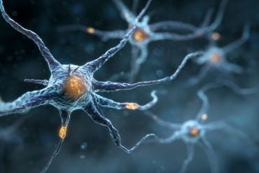
They showed that too much inflammation in the womb impairs the maturation of nerve cells (called neurons), leading to reduced electric activity.
Neurons are the cells in our brain and spinal cord that receive and transmit electrical signals that control the way we walk, hear, see, and think.
Brain development impacts
Lead researcher, Sharmony Kelly says their findings will have a positive impact in preventing or reducing a wide range of conditions that can affect babies, especially those born preterm.
“Inflammation can lead to cerebral palsy or developmental problems affecting growth, movement, vision and hearing, as well as social and emotional problems, language and cognitive delays and more,” Kelly said.
“By increasing our understanding of exactly how inflammation damages brain development we hope to help prevent these life-long consequences.”
“We also showed that these impairments in nerve cell maturation can be detected using a relatively simple and widely used clinical tool that measures electrical activity in the brain, called electroencephalography,” said Dr. Galinsky.
Delicate balance of brain development
“By improving our understanding of the cellular mechanisms that contribute to inflammation-induced disturbances in brain development, and their functional consequences, we can improve our ability to detect neurodevelopment impairments earlier.”
Sharmony Kelly likened the impact of inflammation to a disruption in the symphony of brain development.
“Imagine the brain as an orchestra playing a melody of growth and connectivity; when it’s exposed to inflammation, like an out-of-tune instrument, the orchestra’s performance becomes distorted.”
“If the delicate balance of brain activity, cellular growth, and immune responses is disrupted, potentially leading to long-lasting consequences.”
“We’re drawing attention to the significance of maintaining a harmonious environment for healthy brain development and the potential consequences when that balance is disrupted,” she said.
About this brain development research news
Author: Rob Galinsky
Source: Hudson Institute of Medical Research
Contact: Rob Galinsky – Hudson Institute of Medical Research
Image: The image is credited to Neuroscience News
Original Research: Open access.
“Progressive inflammation reduces high-frequency EEG activity and cortical dendritic arborisation in late gestation fetal sheep” by Rob Galinsky et al. Journal of Neuroinflammation
Abstract
Progressive inflammation reduces high-frequency EEG activity and cortical dendritic arborisation in late gestation fetal sheep
Background
Antenatal infection/inflammation is associated with disturbances in neuronal connectivity, impaired cortical growth and poor neurodevelopmental outcomes. The pathophysiological substrate that underpins these changes is poorly understood. We tested the hypothesis that progressive inflammation in late gestation fetal sheep would alter cortical neuronal microstructure and neural function assessed using electroencephalogram band power analysis.
Methods
Fetal sheep (0.85 of gestation) were surgically instrumented for continuous electroencephalogram (EEG) recording and randomly assigned to repeated saline (control; n = 9) or LPS (0 h = 300 ng, 24 h = 600 ng, 48 h = 1200 ng; n = 8) infusions to induce inflammation. Sheep were euthanised 4 days after the first LPS infusion for assessment of inflammatory gene expression, histopathology and neuronal dendritic morphology in the somatosensory cortex.
Results
LPS infusions increased delta power between 8 and 50 h, with reduced beta power from 18 to 96 h (P < 0.05 vs. control). Basal dendritic length, numbers of dendritic terminals, dendritic arborisation and numbers of dendritic spines were reduced in LPS-exposed fetuses (P < 0.05 vs. control) within the somatosensory cortex. Numbers of microglia and interleukin (IL)-1β immunoreactivity were increased in LPS-exposed fetuses compared with controls (P < 0.05). There were no differences in total numbers of cortical NeuN + neurons or cortical area between the groups.
Conclusions
Exposure to antenatal infection/inflammation was associated with impaired dendritic arborisation, spine number and loss of high-frequency EEG activity, despite normal numbers of neurons, that may contribute to disturbed cortical development and connectivity.


