

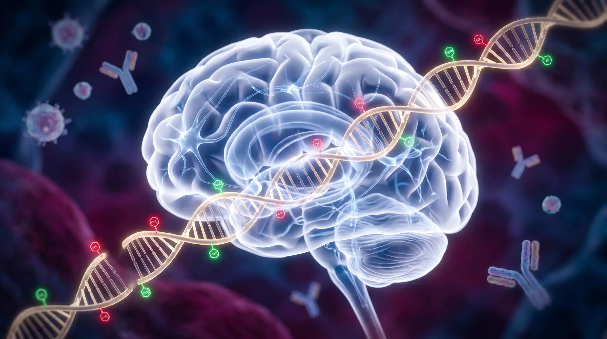
Summary: Researchers have discovered that maternal immune activation during pregnancy alters the epigenome of fetal brain cells, disrupting deep-layer neurons and increasing the risk of neurodevelopmental disorders like autism.
Key Facts:
- Epigenetic Alterations: Maternal immune activation (MIA) changes the methylation patterns in the fetal frontal cortex, actively blocking the transcription factor Tbr1, which is critical for defining deep-layer neurons.
- Autism Overlap: Approximately 25% of high-confidence genes linked to autism spectrum disorder (found in the SFARI Gene Database) were dysregulated in the deep-layer neurons of MIA-affected offspring.
- Lasting Brain Circuitry Changes: The prenatal immune challenge not only altered fetal gene activity but resulted in impaired neurodevelopment and electrophysiological changes that persisted into adulthood.
Source: Salk Institute
Neurodevelopmental conditions, including autism spectrum disorders and ADHD, affect approximately ten percent of the U.S. population. While pinpointing the exact origins of these conditions remains challenging, researchers have long observed a compelling pattern: severe maternal illness during pregnancy correlates with a higher risk of neurodevelopmental disorders in offspring.
Now, scientists at the Salk Institute have uncovered the biological mechanisms behind this phenomenon. By analyzing the epigenomes of mouse frontal cortex cells throughout fetal development, the team discovered thousands of epigenetic differences between offspring from healthy pregnancies and those from pregnancies affected by maternal immune activation.
The study, recently published in Molecular Psychiatry, focused on the epigenome—the chemical tags and modifications layered on top of our base genetic code that dictate which genes are expressed.
Decades ago, epidemiologists noticed that influenza infections during the second or third trimester increased the incidence of psychiatric disorders in children. More recent research linked this to elevated levels of maternal IL-6, an inflammatory protein deployed during the immune response. To explore this, the Salk team utilized a well-characterized mouse model treated with viral mimetic Poly(I:C), which safely mimics an influenza infection and triggers maternal immune activation (PIC-MIA).
The team analyzed mouse neurons in the frontal cortex from mid-gestation to two weeks post-birth. They found that mice from PIC-MIA pregnancies exhibited distinct changes in both gene activity and methylation—a process where small chemical tags attach to DNA to modify its function.
These methylation patterns were heavily concentrated in genomic areas responsible for forming deep-layer neurons. Specifically, genomic sites where the transcription factor Tbr1 typically binds were hypermethylated. Even though there was plenty of Tbr1 protein present—a major regulator for developing brains—the excess methylation blocked it from binding correctly. Consequently, the genomic areas Tbr1 interacts with were downregulated, impairing the proper development of deep-layer neurons.
“We compared our findings to the SFARI Gene Database, an established database of autism spectrum-associated genomic alterations,” explained Jessica Arzavala, co-first author of the study and graduate researcher at Salk. “Among high-confidence genes—those we are most sure are correctly mapped and linked to autism spectrum disorder—around 25% of the database was also dysregulated in our dataset.”
Subsequent electrophysiological recordings of the offspring confirmed that deep-layer neuron development was profoundly impaired, creating lasting changes in brain circuitry that carried into adulthood.
“Infection changes the odds of whether neurodevelopment is affected—not everyone who gets sick during pregnancy is going to definitively have a child with a neurodevelopmental disorder,” cautioned co-corresponding author Dr. Joseph Ecker, a professor at Salk and Howard Hughes Medical Institute investigator.
The study represents a crucial step forward in understanding the cascading consequences of maternal infection. By mapping these exact epigenetic roadblocks, researchers hope to eventually develop maternal or fetal therapeutics capable of mitigating or preventing the risks of neurodevelopmental disorders.
“It’s just the tip of the iceberg,” added co-corresponding author Dr. Margarita Behrens. “Now we can approach questions with more detail. It’s going to be a lot of fun moving forward.”
Editorial Notes:
- This article was edited by a Neuroscience News editor.
- Journal paper reviewed in full.
- Additional context added by our staff.
About this Neurodevelopment Research:
- Media Contact: Isabella Davis
- Source: Salk Institute
- Image Credit: Image generated for Neuroscience News
- Original Research is Open Access: Molecular Psychiatry (September 2, 2026). “Poly(I:C) maternal immune activation alters epigenetic programming in the developing frontal cortex.” Authors: Chi-Yu Lai, Jessica Arzavala, Antonio Pinto-Duarte, Hanqing Liu, Julia Osteen, Rosa Gomez Castanon, Joseph Nery, Shiyuan Wang, Junhao Li, Susan B. Powell, Eran Mukamel, Margarita Behrens, and Joseph Ecker.
- DOI: 10.1038/s41380-026-03856-1
Abstract
Lai, CY., Arzavala, J., Pinto-Duarte, A. et al. Maternal immune activation disrupts epigenomic and functional maturation of cortical excitatory neurons. Mol Psychiatry (2026). https://doi.org/10.1038/s41380-026-03856-1
Elevated levels of maternal pro-inflammatory cytokines following severe infection during gestation can disrupt offspring neural development and increase the risk of neurodevelopmental disorders. The viral mimetic Poly(I:C) reproduces the effects of gestational influenza exposure, leading to behavioral outcomes that recapitulate neurodevelopmental disorder phenotypes. Although Poly(I:C)-induced maternal immune activation (PIC-MIA) alters the epigenome, behavior and cognition of offspring in adulthood, it remains unclear when these changes occur and how MIA influences the epigenomic regulatory programming across the transition from embryonic development to the mature brain. Here, we examined the effects of PIC-MIA on the epigenomic maturation of the frontal cortex, focusing on excitatory neuron-specific DNA methylation and transcriptomic dynamics throughout perinatal development. Mid-gestation PIC-MIA disrupted development of the excitatory neuron transcriptome, with the largest alterations observed at birth. PIC-MIA altered the development of the mature DNA methylation program of excitatory neurons at thousands of genomic regulatory regions that normally gain or lose methylation during development. Transcription factor binding site analyses of these differentially methylated regions revealed a significant enrichment of Tbr1 motifs within hyper-methylated deep-layer neuron-specific regions at birth. Notably, transcriptional targets of Tbr1 were down-regulated at birth despite up-regulation of Tbr1 transcription, suggesting PIC-MIA uncouples Tbr1 expression from its regulatory function in deep-layer neurons. Electrophysiological recordings of intrinsic and firing properties further confirmed a lasting disruption in deep-layer neuronal activity. Our results suggest that mid-gestation MIA may alter the development of deep-layer neurons through an epigenomic blockade of Tbr1 function, thereby perturbing normal cortical circuit formation.


