

Two parts of hippocampus work together to determine whether stimulus is completely new or related to something familiar.
You see a man at the grocery store. Is that the guy you went to college with or just someone who looks like him?
One tiny spot in the brain has the answer.
Johns Hopkins University neuroscientists have identified the part of the hippocampus that creates and processes this type of memory, furthering our understanding of how the mind works, and what’s going wrong when it doesn’t. Their findings are published in the current issue of the journal Neuron.
“You see a familiar face and say to yourself, ‘I think I’ve seen that face.’ But is this someone I met five years ago, maybe with thinner hair or different glasses—or is it someone else entirely,” said James J. Knierim, a professor of neuroscience at the university’s Zanvyl Krieger Mind/Brain Institute who led the research. “That’s one of the biggest problems our memory system has to solve.”
Neural activity in the hippocampus allows someone to remember where they parked their car, find their home even if the paint color changes, and recognize an old song when it comes on the radio.
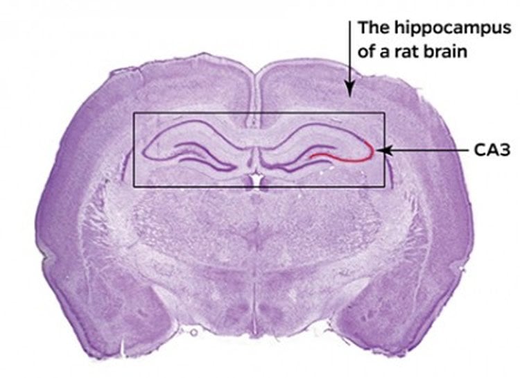
Brain researchers theorized that two parts of the hippocampus—the dentate gyrus and CA3—compete to decide whether a stimulus is completely new or an altered version of something familiar. The dentate gyrus was thought to automatically encode each stimulus as new, a process called pattern separation. In contrast, CA3 was thought to minimize any small changes from one experience to the next and classify the stimuli as being the same, a process called pattern completion. So, the dentate gyrus would assume that the person with thinner hair and unfamiliar glasses was a complete stranger, while CA3 would ignore the altered details and retrieve the memory of a college buddy.
Prior work by Knierim’s group and others provided evidence in favor of this long-standing theory. The new research shows, however, that CA3 is more complicated than previously thought—parts of CA3 come to different decisions, and they pass these different decisions to other brain areas.
“The final job of the CA3 region is to make the decision: Is it the same or is it different?” Knierim said. “Usually you are correct in remembering that this person is a slightly different version of the person you met years ago. But when you are wrong, and it embarrassingly turns out that this is a complete stranger, you want to create a memory of this new person that is absolutely distinct from the memory of your familiar friend, so you don’t make the mistake again.”
Knierim and Johns Hopkins postdoctoral fellows Heekyung Lee and Cheng Wang, along with Sachin S. Deshmukh, a former assistant research scientist in Knierim’s lab, monitored rats as they got to know an environment and as that environment changed.
The team implanted electrodes in the hippocampus of the rats. They trained the rats to run around a track, eating chocolate sprinkles. The track floor had four different textures—sandpaper, carpet padding, duct tape and a rubber mat. The rat could see, feel and smell the differences in the textures. Meanwhile, a black curtain surrounding the track had various objects attached to it. Over 10 days, the rats built mental maps of that environment.
Then the experimenters changed things up. They rotated the track counter-clockwise, while rotating the curtain clockwise, creating a perceptual mismatch in the rats’ minds. The effect was similar, Knierim said, to if you opened the door of your home and all of your pictures were hanging on different walls and your furniture had been moved.
“Would you recognize it as your home or think you are lost?” he said. “It’s a very disorienting experience and a very uncomfortable feeling.”
Even when the perceptual mismatch between the track and curtain was small, the “pattern separating” part of CA3 almost completely changed its activity patterns, creating a new memory of the altered environment. But the “pattern completing” part of CA3 tended to retrieve a similar activity pattern used to encode the original memory, even when the perceptual mismatch increased.
The findings, which validate models about how memory works, could help explain what goes wrong with memory in diseases like Alzheimer’s and could help to preserve people’s memories as they age.
Funding: This research was supported by the National Institute of Health grants R01 NS039456 and R01 MH094146 and by the Johns Hopkins University Brain Sciences Institute.
Source: Jill Rosen – Johns Hopkins University
Image Source: The image is credited to Johns Hopkins University
Original Research: Abstract for “Neural Population Evidence of Functional Heterogeneity along the CA3 Transverse Axis: Pattern Completion versus Pattern Separation” by Heekyung Lee, Cheng Wang, Sachin S. Deshmukh, and James J. Knierim in Neuron. Published online August 19 2015 doi:10.1016/j.neuron.2015.07.012
Abstract
Neural Population Evidence of Functional Heterogeneity along the CA3 Transverse Axis: Pattern Completion versus Pattern Separation
Highlights
•Neural population analyses reveal functional dissociation along CA3 transverse axis
•Proximal CA3 population activity demonstrates computational pattern separation
•Distal CA3 population activity demonstrates computational pattern completion
•CA2 population activity is similar to distal CA3 in a cue-mismatch environment
Summary
Classical theories of associative memory model CA3 as a homogeneous attractor network because of its strong recurrent circuitry. However, anatomical gradients suggest a functional diversity along the CA3 transverse axis. We examined the neural population coherence along this axis, when the local and global spatial reference frames were put in conflict with each other. Proximal CA3 (near the dentate gyrus), where the recurrent collaterals are the weakest, showed degraded representations, similar to the pattern separation shown by the dentate gyrus. Distal CA3 (near CA2), where the recurrent collaterals are the strongest, maintained coherent representations in the conflict situation, resembling the classic attractor network system. CA2 also maintained coherent representations. This dissociation between proximal and distal CA3 provides strong evidence that the recurrent collateral system underlies the associative network functions of CA3, with a separate role of proximal CA3 in pattern separation.
“Neural Population Evidence of Functional Heterogeneity along the CA3 Transverse Axis: Pattern Completion versus Pattern Separation” by Heekyung Lee, Cheng Wang, Sachin S. Deshmukh, and James J. Knierim in Neuron. Published online August 19 2015 doi:10.1016/j.neuron.2015.07.012


