Summary: A new study pinpoints he areas of the brain involved in decision-making and action.
Source: Johns Hopkins University.
Johns Hopkins University researchers are the first to glimpse the human brain making a purely voluntary decision to act.
Unlike most brain studies where scientists watch as people respond to cues or commands, Johns Hopkins researchers found a way to observe people’s brain activity as they made choices entirely on their own. The findings, which pinpoint the parts of the brain involved in decision-making and action, are now online, and due to appear in a special October issue of the journal Attention, Perception, & Psychophysics.
“How do we peek into people’s brains and find out how we make choices entirely on our own?” asked Susan Courtney, a professor of psychological and brain sciences. “What parts of the brain are involved in free choice?”
The team devised a novel experiment to track a person’s focus of attention without using intrusive cues or commands. Participants, positioned in MRI scanners, were left alone to watch a split screen as rapid streams of colorful numbers and letters scrolled past on each side. They were asked simply to pay attention to one side for a while, then to the other side — when to switch sides was entirely up to them. Over an hour, the participants switched their attention from one side to the other dozens of times.
Researchers monitored the participants’ brains as they watched the media stream, both before and after they switched their focus.
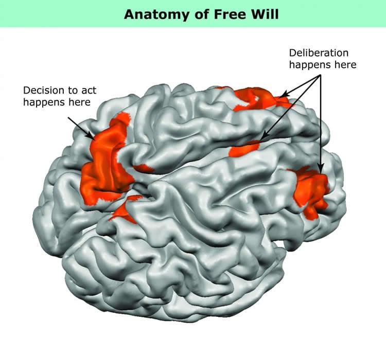
For the first time, researchers were able to see both what happens in a human brain the moment a free choice is made, and what happens during the lead-up to that decision — how the brain behaves during the deliberation over whether to act.
The actual switching of attention from one side to the other was closely linked to activity in the parietal lobe, near the back of the brain. The activity leading up to the choice — that is, the period of deliberation — occurred in the frontal cortex, in areas involved in reasoning and movement, and in the basal ganglia, regions deep within the brain that are responsible for a variety of motor control functions including the ability to start an action. The frontal-lobe activity began earlier than it would have if participants had been told to shift attention, clearly demonstrating that the brain was preparing a purely voluntary action rather than merely following an order.
Together, the two brain regions make up the core components underlying the will to act, the authors concluded.
“What’s truly remarkable about this project,” said Leon Gmeindl, a research scientist at Johns Hopkins and lead author of the study, “is that by devising a way to detect brain events that are otherwise invisible — that is, a kind of high-tech ‘mind reading’ — we uncovered important information about what may be the neural underpinnings of volition, or free will.”
Now that scientists have a way to track choices made from free will, they can use the technique to determine what’s happening in the brain as people wrestle with other, more complex decisions. For instance, researchers could observe the brain as someone tried to decide between snacking on a doughnut or on an apple — watching as someone weighed short-term rewards against long-term rewards, and perhaps being able to pinpoint the tipping point between the two.
“We now have the ability to learn more about how we make decisions in the real world,” Courtney said.
The research team also included former Johns Hopkins doctoral students and postdoctoral fellows Yu-Chin Chiu, Michael S. Esterman, and Adam S. Greenberg. The paper is dedicated to the last author of the study, Steven Yantis, a professor in the Department of Psychological and Brain Sciences who died of cancer in 2014.
Funding: This research was supported by National Institutes of Health grants T32EY07143, T32AG027668, F31-NS055664, R01-MH082957, and R01-DA013165.
Source: Johns Hopkins University
Image Source: This NeuroscienceNews.com image is credited to Johns Hopkins University.
Video Source: The video is credited to Johns Hopkins University.
Original Research: Abstract for “Tracking the will to attend: Cortical activity indexes self-generated, voluntary shifts of attention” by Leon Gmeindl, Yu-Chin Chiu , Michael S. Esterman, Adam S. Greenberg, Susan M. Courtney, and Steven Yantis in Attention, Perception, & Psychophysics. Published online June 14 2016 doi:10.3758/s13414-016-1159-7
[cbtabs][cbtab title=”MLA”]Johns Hopkins University. “What “Free Will” Looks Like in the Brain.” NeuroscienceNews. NeuroscienceNews, 13 July 2016.
<https://neurosciencenews.com/free-will-decision-making-neuroscience-4673/>.[/cbtab][cbtab title=”APA”]Johns Hopkins University. (2016, July 13). What “Free Will” Looks Like in the Brain. NeuroscienceNew. Retrieved July 13, 2016 from https://neurosciencenews.com/free-will-decision-making-neuroscience-4673/[/cbtab][cbtab title=”Chicago”]Johns Hopkins University. “What “Free Will” Looks Like in the Brain.” https://neurosciencenews.com/free-will-decision-making-neuroscience-4673/ (accessed July 13, 2016).[/cbtab][/cbtabs]
Abstract
Tracking the will to attend: Cortical activity indexes self-generated, voluntary shifts of attention
The neural substrates of volition have long tantalized philosophers and scientists. Over the past few decades, researchers have employed increasingly sophisticated technology to investigate this issue, but many studies have been limited considerably by their reliance on intrusive experimental procedures (e.g., abrupt instructional cues), measures of brain activity contaminated by overt behavior, or introspective self-report techniques of questionable validity. Here, we used multivoxel pattern time-course analysis of functional magnetic resonance imaging data to index voluntary, covert perceptual acts—shifts of visuospatial attention—in the absence of instructional cues, overt behavioral indices, and self-report. We found that these self-generated, voluntary attention shifts were time-locked to activity in the medial superior parietal lobule, supporting the hypothesis that this brain region is engaged in voluntary attentional reconfiguration. Self-generated attention shifts were also time-locked to activity in the basal ganglia, a novel finding that motivates further research into the role of the basal ganglia in acts of volition. Remarkably, prior to self-generated shifts of attention, we observed early and selective increases in the activation of medial frontal (dorsal anterior cingulate) and lateral prefrontal (right middle frontal gyrus) cortex—activity that likely reflects processing related to the intention or preparation to reorient attention. These findings, which extend recent evidence on freely chosen motor movements, suggest that dorsal anterior cingulate and lateral prefrontal cortices play key roles in both overt and covert acts of volition, and may constitute core components of a brain network underlying the will to attend.
“Tracking the will to attend: Cortical activity indexes self-generated, voluntary shifts of attention” by Leon Gmeindl, Yu-Chin Chiu , Michael S. Esterman, Adam S. Greenberg, Susan M. Courtney, and Steven Yantis in Attention, Perception, & Psychophysics. Published online June 14 2016 doi:10.3758/s13414-016-1159-7