

Summary: A new study puts into question conventional belief that the eyes communicate with the brain exclusively via one signaling pathway. Researchers have identified a subset of retinal neurons that sends inhibitory signals to the brain. This subset of neurons is also involved in the synchronization of circadian rhythms to light/dark cycles and pupil constriction to bright light intensity.
Source: Northwestern University
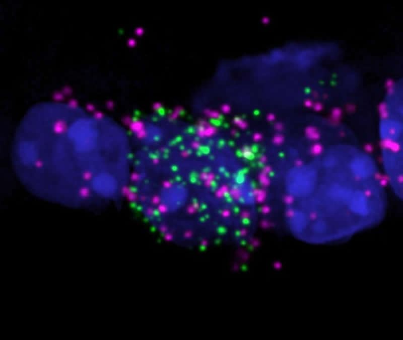
The eyes have a surprise.
For decades, biology textbooks have stated that eyes communicate with the brain exclusively through one type of signaling pathway. But a new discovery shows that some retinal neurons take a road less traveled.
New research, led by Northwestern University, has found that a subset of retinal neurons sends inhibitory signals to the brain. Before, researchers believed the eye only sends excitatory signals. (Simply put: Excitatory signaling makes neurons to fire more; inhibitory signaling makes neurons to fire less.)
The Northwestern researchers also found that this subset of retinal neurons is involved in subconscious behaviors, such as synchronization of circadian rhythms to light/dark cycles and pupil constriction to intense bright lights. By better understanding how these neurons function, researchers can explore new pathways by which light influences our behavior.
“These inhibitory signals prevent our circadian clock from resetting to dim light and prevent pupil constriction in low light, both of which are adaptive for proper vision and daily function,” said Northwestern’s Tiffany Schmidt, who led the research. “We think that our results provide a mechanism for understanding why our eye is so exquisitely sensitive to light, but our subconscious behaviors are comparatively insensitive to light.”
The research will be published in the May 1 issue of the journal Science.
Schmidt is an assistant professor of neurobiology at Northwestern’s Weinberg College of Arts and Sciences. Takuma Sonoda, a former Ph.D. student in the Northwestern University Interdepartmental Neuroscience program, is the paper’s first author.
To conduct the study, Schmidt and her team blocked the retinal neurons responsible for inhibitory signaling in a mouse model. When this signal was blocked, dim light was more effective at shifting the mice’s circadian rhythms.
“This suggests that there is a signal from the eye that actively inhibits circadian rhythms realignment when environmental light changes, which was unexpected,” Schmidt said. “This makes some sense, however, because you do not want to adjust your body’s entire clock for minor perturbations in the environmental light/dark cycle, you only want this massive adjustment to take place if the change in lighting is robust.”
Schmidt’s team also found that, when the inhibitory signals from the eye were blocked, mice’s pupils were much more sensitive to light.
“Our working hypothesis is that this mechanism keeps pupils from constricting in very low light,” Sonoda said. “This increases the amount of light hitting your retina, and makes it easier to see in low light conditions. This mechanism explains, in least part, why your pupils avoid constricting until bright light intensifies.”
Funding: The research, “A non-canonical inhibitory circuit dampens behavioral sensitivity to light,” was supported by a Klingenstein-Simons Fellowship in the Neurosciences, the Alfred P. Sloan Foundation and the National Institutes of Health (award numbers 1DP2EY022584, T32 EY025202 and F31 EY030360-01).
About this neuroscience research article
Source:
Northwestern University
Media Contacts:
Amanda Morris – Northwestern University
Image Source:
The image is credited to Northwestern University.
Original Research: The study will appear in Science.
Feel Free To Share This Visual Neuroscience News.


