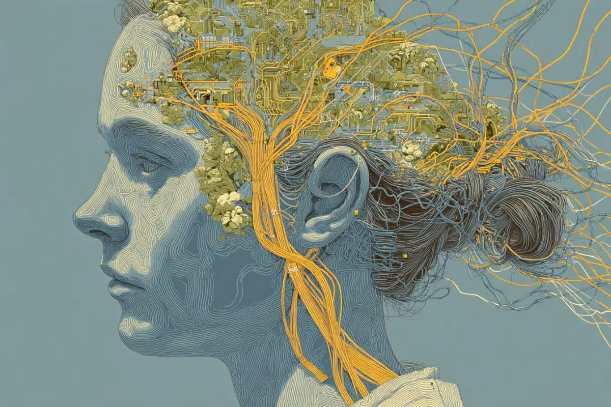
Summary: How do the synchronized rhythms of the human brain emerge during development? While EEGs provide a window into these brain waves, they can’t tell us what’s happening at a cellular level when rhythms go awry in conditions like epilepsy or autism. A new study introduces a simplified, scalable 2D human neuron platform that finally bridges this gap.
By growing networks of human neurons from stem cells (iPSCs) on sensor-embedded plates, researchers successfully recorded the emergence of “nested oscillations”—complex waves within waves that mirror those seen in a developing brain. This high-throughput model allows scientists to systematically test how drugs and genetic mutations reshape brain activity, offering a powerful new tool for neurodevelopmental research.
Key Facts
- The “Nested” Rhythm: The study captured the emergence of complex oscillations (delta, theta, and alpha) layered within slow waves, mimicking the electrical maturation of a real human brain.
- GABA’s Role: Increasing inhibitory GABAergic neurons caused these rhythms to appear earlier, while blocking GABA signaling reduced them, confirming that “calming” signals are essential for organized brain waves.
- Scalable Testing: Unlike complex 3D organoids, this 2D platform is designed for high-volume drug testing and benchmarking, allowing for extensive dose-response studies.
- Separating Signal from Noise: Using a new analysis framework, researchers can now distinguish actual rhythmic peaks from “broadband” background noise, revealing that the “noise” itself often carries vital biological data.
- Clinical Implications: The platform provides a way to study how potassium channel mutations linked to epilepsy disrupt network-level rhythms, potentially accelerating the discovery of new treatments.
Source: Sanford Burnham Prebys
An EEG (electroencephalogram) is a painless test that uses small sensors placed on the scalp to measure the brain’s electrical activity. It provides a real-time readout of brain “waves”—rhythms generated by large groups of neurons working together—and clinicians use it to look for patterns linked to sleep, seizures and other changes in brain function.
These rhythms change as the brain develops, and disruptions to that process have been linked to conditions ranging from epilepsy to neurodevelopmental and psychiatric disorders. EEG recordings can track these changing rhythms, but they cannot uncover what has occurred within the brain’s cells to cause the altered electrical activity.

To learn what governs the emergence of these rhythmic patterns in humans, researchers need more tools. In a study published on January 24, 2026, in Neurobiology of Disease, researchers at Sanford Burnham Prebys Medical Discovery Institute, with collaborators at the University of California San Diego (UCSD) and BioMarin Pharmaceutical, developed a simplified, scalable human cell model to study how coordinated rhythms emerge and how they respond when neurons are perturbed with chemical compounds.
The team’s approach was to grow two-dimensional (2D) networks of human neurons derived from induced pluripotent stem cells (iPSCs). The scientists recorded the neurons’ activity over time using multi-electrode arrays (MEAs), plates embedded with tiny sensors that can monitor many independent networks in parallel.
Because iPSCs can be generated in the lab from accessible donor cells such as from skin or blood samples, they make it possible to produce large numbers of human neurons from both healthy individuals and patients.
As these 2D networks matured, the researchers observed the emergence of “nested oscillations,” slow waves with faster rhythmic structure layered within them. These oscillations were observed across frequency ranges commonly seen in brain recordings (delta, theta and alpha). They then tested how specific biological mechanisms influence these rhythms.
“The results of these and other experiments show that this simplified 2D neuronal network model captures key features of network maturation, and gives us the scale and control needed for systematic testing,” said Anne Bang, PhD, the study’s senior and co-corresponding author, associate professor in the Center for Therapeutics Discovery at Sanford Burnham Prebys and director of Cell Biology at the Conrad Prebys Center for Chemical Genomics.
The new work positions 2D networks as complementary to three-dimensional brain organoids. Organoids, also produced from iPSCs, can recapitulate aspects of tissue architecture, cellular diversity and network activity that are difficult to reproduce in 2D formats.
At the same time, organoid complexity can make certain experiments harder to run at scale, particularly studies that require large numbers of replicates or extensive dose–response testing across many conditions.
“Organoids are invaluable for modeling aspects of brain organization,” said Bang. “What we add here is a complementary 2D platform that emphasizes experimental control and throughput, capabilities that can be especially useful for benchmarking and systematic testing in disease modeling and early-stage therapeutic evaluation.”
One major focus of the team’s study was inhibitory signaling mediated by GABA, a neurotransmitter produced by GABAergic neurons that helps stabilize and calm network activity. For instance, this inhibitory system helps promote sleep and prevent seizures.
The team found that nested rhythms were reduced when GABA signaling was blocked with a GABA-A receptor antagonist drug, and that increasing the proportion of GABAergic neurons in the network caused these rhythms to emerge earlier.
Their findings align with prior evidence in the field and support follow-up studies on how GABA-mediated inhibition shapes the emergence and maturation of oscillations in iPSC-based models of neurodevelopmental and psychiatric disease.
The researchers also tested drugs that affect potassium channels. These proteins help set a neuron’s ability to generate and transmit electrical signals, which is known as neuronal excitability.
“We wanted to explore potassium channels because certain mutant forms have been implicated in neurological disorders such as epilepsy and neurodevelopmental syndromes,” said Bang.
Their results suggested that different potassium channel perturbations can influence rhythmic organization in distinct ways, highlighting that excitability is not a single dial that can easily be turned up or down, and that specific mechanisms may have specific network-level signatures.
To strengthen interpretation of these recordings, the team used analysis methods developed in the laboratory of Bradley Voytek, PhD, co-corresponding author and professor and chair of Cognitive Sciences at UCSD.
This framework separates two components of neural signals: oscillations (rhythmic peaks) and a broadband background signal that is often treated as random “noise” that lacks useful data.
In some experiments in the new study, the broadband component shifted alongside oscillatory measures, suggesting it is not just background noise but can carry biologically meaningful information about the network. Measuring both components helps determine whether a drug is changing a specific rhythm, shifting the overall network state or both.
Finally, the study evaluated a faster neuron-production method based on inducing expression of the transcription factor NEUROG2 (NGN2) in iPSCs. These NGN2-induced networks showed only rudimentary nested rhythms, suggesting that rapid differentiation approaches may require additional optimization to reliably capture rhythmic features.
“Improving these faster methods to generate neurons could broaden what is possible in higher-throughput settings,” said Bang.
By combining scalable human 2D neuronal networks with analysis that distinguishes rhythmic peaks from the broadband background, the research team’s approach provides a practical way to study how coordinated activity emerges and to test how specific pathways or drug-mediated perturbations reshape network dynamics.
Over time, this kind of controlled, reproducible platform can help build reference benchmarks for comparing genetic backgrounds, disease models and candidate treatments.
Funding: The study was supported by the National Institutes of Health, National Institute of Mental Health, National Institute of General Medical Sciences, BioMarin Pharmaceutical, Viterbi Family Foundation of the Jewish Community Foundation San Diego and Burroughs Wellcome Fund.
Key Questions Answered:
A: 3D organoids are great for structure, but they are hard to replicate consistently in large numbers. These “flat” 2D networks are like a high-speed testing lab—they give scientists the control and volume needed to test thousands of drug doses and genetic variations quickly.
A: Think of them as a rhythm within a rhythm. In the brain, these complex patterns are a sign of maturity. When these layers don’t form correctly, it can lead to seizures or developmental delays. Being able to grow and watch these layers form in a dish is a huge leap forward.
A: Yes. By using skin or blood cells from patients with these conditions to create the neurons, scientists can see exactly how their specific “rhythms” differ from healthy brains and test which drugs might “tune” their brain activity back to normal.
Editorial Notes:
- This article was edited by a Neuroscience News editor.
- Journal paper reviewed in full.
- Additional context added by our staff.
About this neuroscience research news
Author: Greg Calhoun
Source: Sanford Burnham Prebys
Contact: Greg Calhoun – Sanford Burnham Prebys
Image: The image is credited to Neuroscience News
Original Research: Open access.
“Pharmacological manipulation of nested oscillations in human iPSC-derived 2D neuronal networks” by Deborah Pré, Christian Cazares, Alexander T. Wooten, Haowen Zhou, Isabel Onofre, Ashley Neil, Todd Logan, Ruilong Hu, Jan H. Lui, Bradley Voytek, and Anne G. Bang. Neurobiology of Disease
DOI:10.1016/j.nbd.2026.107281
Abstract
Pharmacological manipulation of nested oscillations in human iPSC-derived 2D neuronal networks
Dynamically coupled neural networks are fundamental to human cognition and behavior and are disrupted in neurodevelopmental disorders. The formation and dissolution of functional networks is thought to be driven by synchronized oscillatory bursts across large populations of neurons.
The mechanisms driving the emergence of these rhythms, known as oscillogenesis, are not well understood, particularly in the human brain. Using multi-electrode arrays, we investigated oscillogenesis in human induced pluripotent stem cell 2D neural cultures at different developmental stages and under pharmacological challenges.
We found that cultures exhibited nested oscillations that were reduced by GABAA receptor blockade and emerged earlier when the proportion of GABAergic neurons was increased. Pharmacological manipulations of voltage-gated potassium channels and cholinergic receptors modulated the pattern of nested oscillations.
These results reveal the capacity of these 2D cultures to model oscillogenesis, and underscore the need for their continued refinement, paving the way for linking systems-level neural networks to human cognition and disease.