Summary: While both the flu and COVID-19 are respiratory viruses that can leave lasting lung damage, only COVID-19 appears to leave a persistent, inflammatory footprint on the brain. New research reveals that SARS-CoV-2 causes small blood vessel injury and ongoing brain inflammation even after the virus is cleared.
This study found that COVID-19 specifically disrupts serotonin and dopamine signaling—neurotransmitters critical for mood, cognition, and energy. These findings provide a biological explanation for why “brain fog” and fatigue are hallmark symptoms of Long COVID, but are rarely seen after the seasonal flu.
Key Facts
- Vascular Injury: COVID-19, but not the flu, causes tiny areas of bleeding and inflammation in the brain’s small blood vessels, even in mild cases.
- Neurotransmitter Disruption: SARS-CoV-2 infection alters gene expression related to serotonin and dopamine regulation, which explains the persistent cognitive and mood changes in Long COVID.
- Lung Repair Failure: While both viruses cause lung scarring (collagen buildup), the lungs switch into “repair mode” after the flu. Following COVID-19, this natural healing response is largely missing.
- Absence of Virus: Strikingly, the brain inflammation occurred despite the fact that no actual virus was detected in the brain tissue, suggesting the damage is caused by the body’s overactive immune response.
- Subchronic Effects: The study focused on the “subchronic” phase—weeks after the initial infection—to specifically map out the transition from acute illness to long-term symptoms.
Source: Tulane University
Even a mild case of COVID-19 or the flu can impact the body long after the fever and cough fade, according to new Tulane University research that may help explain why some people struggle to feel fully recovered weeks or months later.
Tulane researchers found that while both viruses can leave lasting lung damage, only SARS-CoV-2 infection caused persistent brain inflammation and small blood vessel injury, even after the virus was no longer detectable.

The findings, published in Frontiers in Immunology, help explain why long COVID often includes neurological symptoms such as brain fog, fatigue and mood changes, while influenza is more commonly associated with respiratory complications.
“Influenza and COVID-19 affect large populations worldwide and carry a significant public health toll, yet the mechanisms behind their long-term effects remain poorly understood,” said Dr. Xuebin Qin, lead author and professor of microbiology and immunology at the Tulane National Biomedical Research Center.
To separate effects common to severe respiratory infections from those unique to COVID-19, researchers used a mouse model to examine lung and brain tissue after infection had cleared.
In the lungs, both viruses left behind a similar picture: immune cells that failed to fully stand down and increased buildup of collagen, a protein associated with scarring. Those changes can stiffen lung tissue and make breathing feel more labored — a possible biological explanation for why some people report lingering shortness of breath after respiratory infections.
But when the researchers looked more closely, they found a key difference. After the flu, the lungs appeared to switch into repair mode, sending specialized cells into damaged areas to help rebuild the lining of the airways. That repair response was largely missing after COVID-19 infection, suggesting the virus may interfere with the lung’s natural healing process.
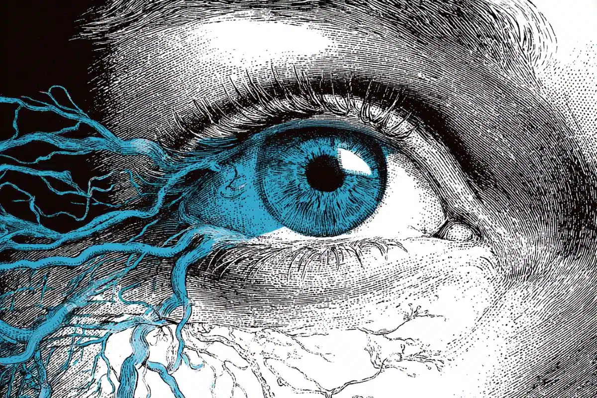
The most striking differences appeared in the brain.
Although neither virus was found in brain tissue, mice that had COVID-19 showed signs of persistent brain inflammation weeks later, along with tiny areas of bleeding. Gene expression analysis revealed ongoing inflammatory signaling and disruption of pathways involved in serotonin and dopamine regulation, systems closely tied to mood, cognition and energy levels. These persistent changes were largely absent in influenza-infected animals.
“In both infections, we observed lasting lung injury,” Qin said. “But long-term effects in the brain were unique to SARS-CoV-2. That distinction is critical to understanding long COVID.”
This study was supported by an American Heart Association award Qin received as part of a national effort to understand the long-term cardiovascular and cerebrovascular effects of COVID-19. The findings shed new light on how vascular and immune changes may contribute to persistent neurological symptoms.
By defining these biological changes, the research offers a clearer foundation for monitoring patients and developing treatments aimed at preventing lasting damage. As lingering symptoms continue to complicate recovery for some, understanding what is driving them is essential to reducing long-term health consequences.
Funding: This research was supported by the American Heart Association Long COVID Impact Project (AHA962950), the National Institutes of Health, including P51OD011104-62 and R01HL165265, and institutional funding.
Key Questions Answered:
A: This study shows that while both can scar your lungs, only COVID-19 effectively “sets fire” to your brain’s communication lines. By damaging small blood vessels and disrupting serotonin and dopamine, COVID creates a biological environment for depression, fatigue, and “fog” that the flu simply doesn’t.
A: It’s an immune “echo.” Even after the virus is gone, your body’s inflammatory signals stay turned on in the brain. This ongoing inflammation causes small leaks in blood vessels and messes with the chemicals that help you think and feel happy.
A: The research found that after the flu, the body sends in a “repair crew” to fix the airway lining. After COVID, that crew never shows up. This suggests we may need specific treatments to jumpstart the healing process in Long COVID patients.
Editorial Notes:
- This article was edited by a Neuroscience News editor.
- Journal paper reviewed in full.
- Additional context added by our staff.
About this COVID-19 and neurology research news
Author: Andrew Yawn
Source: Tulane University
Contact: Andrew Yawn – Tulane University
Image: The image is credited to Neuroscience News
Original Research: Open access.
“Characterization of Subchronic Lung and Brain Consequences Caused by Mouse-Adapted SARS-CoV-2 and Influenza A Infection of C57BL6 mice” by Joshua Currey, Chenxiao Wang, Meredith G. Mayer, Yilin Chen, Ana Karina Nisperuza Vidal, Michaela J. Allen, Mst Shamima Khatun, Calder R. Ellsworth, Mohammad Islamuddin, Jefferson Evangelista, Skye M. Minor, Nadia Golden, Kevin J. Zwezdaryk, Nicholas J. Maness, Robert V. Blair, Jay K. Kolls, Derek A. Pociask, Tracy Fischer, and Xuebin Qin. Frontiers in Immunology
DOI:10.3389/fimmu.2026.1755141
Abstract
Characterization of Subchronic Lung and Brain Consequences Caused by Mouse-Adapted SARS-CoV-2 and Influenza A Infection of C57BL6 mice
Introduction:
SARS-CoV-2 and, to a lesser extent, influenza A can lead to long-term complications in the respiratory and nervous systems. However, the mechanisms driving post-viral sequelae remain poorly understood.
Methods:
To address this gap, we longitudinally characterized C57BL/6 mice infected with sublethal doses of mouse-adapted SARS-CoV-2 (MA30) or influenza A (PR8). Lung and brain tissues were analyzed at 14-, 21-, and 28-days post-infection (DPI) using histological analysis and bulk-RNA sequencing.
Results:
In the lungs, both infections caused prolonged inflammation and fibrosis. MA30-infected lungs showed persistent upregulation of inflammation, coagulation, complement, as well as fibrotic, and extracellular matrix (ECM) remodeling pathways at 21 DPI, alongside downregulation of epithelial junction and metabolic program pathways.
In contrast, PR8-infected lungs exhibited a strong acute interferon response and chronic upregulation of basal epithelial markers (e.g., Krt5, Krt14), consistent with epithelial regeneration. Notably, only PR8-infected mice displayed KRT5+ progenitor cell migration into damaged lung regions, indicating divergence in repair mechanisms.
Neither MA30-infected, nor PR8-infected mice had detectable brain infection. However, MA30 mice, but not PR8-infected mice exhibited an elevated frequency of microhemorrhages at early timepoints and marked neuroinflammation at all timepoints. Transcriptomic profiling of MA30-infected brains showed enrichment for up-regulation of ECM remodeling, vascular dysfunction, IL6-signaling pathways along with a virus-specific disruption of the hypothalamic–pituitary axis with MA30 infection not seen in PR8-infected brains.
These included genes linked to neuroinflammation, sensory processing disruption, and microvascular injury, mirroring clinical features of Long COVID.
Discussion:
Together, these findings establish distinct tissue-specific trajectories of long-term pathology following SARS-CoV-2 and influenza infection and provide a foundation for dissecting the mechanisms of post-viral lung and brain disease.