

Summary: Researchers have successfully mapped brain to spinal cord neural connections that help drive voluntary movement. The map could be beneficial in developing new strategies to treat spinal cord damage.
Source: Cincinnati Children’s Hospital Medical Center.
Researchers trying to help people suffering from paralysis after a spinal cord injury or stroke mapped critical brain-to-spinal cord nerve connections that drive voluntary movement in forelimbs, a development that scientists say allows them to start looking for specific repair strategies.
The study by Yutaka Yoshida, PhD, and colleagues at Cincinnati Children’s Hospital Medical Center is an important step toward rehabilitating motor circuits to help motor function recover after an injury or disease damages the central nervous system, the scientists report in Cell Reports.
“The map described in this study should allow us to explore which corticospinal-spinal interneuron connections are good targets for repair and restoration of voluntary movement,” says Yoshida, lead investigator in the Division of Developmental Biology. “More research is necessary before human therapies are possible, but this information is very helpful for future repair strategies. We now know which circuits need to be repaired.”
The scientists said it will take years for additional investigative work to make the current findings therapeutically relevant. Yoshida and colleagues are conducting new studies to build on the basic neuronal architecture identified in the current study. They want to reach a point where these circuits can be reconstructed to stimulate the recovery of motor function central nervous system injuries.
Corticospinal Schematics
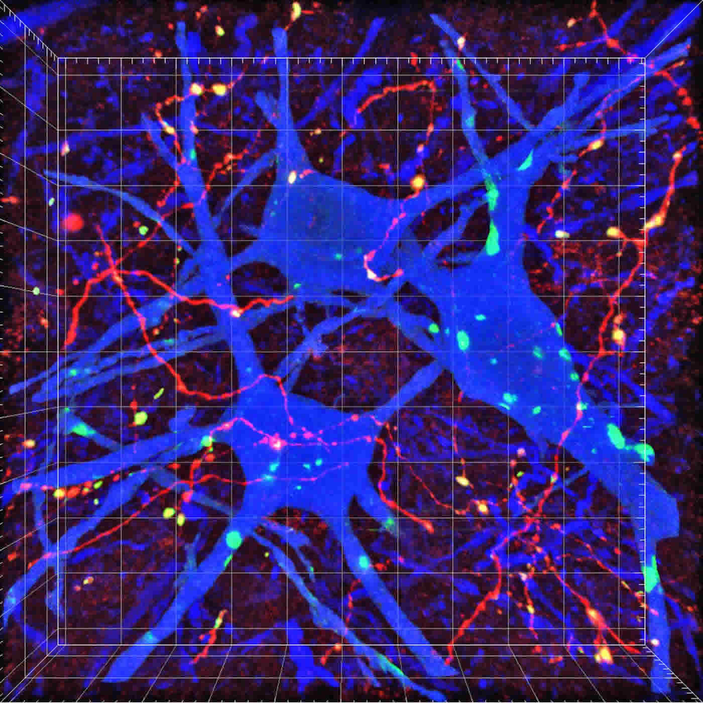
Little has been known about how the corticospinal network of nerve connections between the brain and spinal cord are organized and function together. Seemingly simple tasks like reaching or grabbing require precise coordination between sensory and motor information transmitted through these coordinated connections, according to the researchers.
To map this connectivity in the current study, the scientists study these circuits in laboratory mice–taking advantage of similar corticospinal connections in primates, cats, and rodents.
Working initially from previous studies by his research team and others, Yoshida and colleagues were able to track corticospinal connections from the brain’s cerebral cortex near the top of the head down to the spinal cord. They also traced the organization and function of corticospinal circuits using mouse genetics, and a viral tracer (a de-armed rabies virus) that allowed investigators to highlight and capture images of these links.
The connections trace down through what’s called the brain’s internal capsule, then arrive at the caudal medulla of the brain just above the spinal cord. From there they enter the spinal cord, crisscrossing deep inside the spine as they continue to protrude downward and make additional connections.
Yoshida said his team was able to develop a map of corticospinal neurons that control forelimb and sensory nerve impulses. They also identified specific neurons that control different skilled movements.
In these areas, the scientists show how the nerve fibers connect onto certain premotor interneurons and transmit impulses between neurons to trigger skilled movements. This includes nerve fibers that express a transcription factor called Chx10 (a regulator gene that instructs other genes to turn on or off to initiate biological functions).
Chx10 is linked to nervous system function in other parts of the body, including the eyes. When the researchers silenced Chx10 only in the cervical spinal cord, it hampered the animals’ ability to reach for food.
The Importance of Sensing
The researchers also highlighted the connections of corticospinal neurons in the forelimb sensory cortex–which control the animals’ ability to sense and convert external stimuli into electrical impulses. They said that in contrast to corticospinal neurons in the motor cortex that directly trigger certain skilled movement, corticospinal neurons in the sensory cortex do not connect directly to premotor neurons. Instead, they connect directly to other spinal interneurons that express a gene called Vglut3.
This is important because when the scientist inhibited neurons expressing Vglut3 in the cervical spinal cord, it also caused deficits in the animals’ ability to grab and release food pellets, as well as other goal-oriented tasks.
Key collaborators on this study include scientists at the Precursory Research for Embryonic Science and Technology (PRESTO) at the Japan Science and Technology Agency, the Brain Research Institute at Niigata University in Japan and the University of Cincinnati Medical Center.
Funding: Funding support for the research came in part from: the National Institute of Neurological Disorders and Stroke (NS093002) PRESTO (JST-JPMJPR13M8), the Japan Society for the Promotion of Science (JSPS) Grants-in-Aid for Scientific Research (KAKENHI 17H04985, 17H05556, 17K19443); a JSPS Postdoctoral Fellowships for Research Abroad, the Foundation for the Promotion of Medical Science, Kato Memorial Bioscience Foundation, Grant-in-Aid from the Tokyo Biochemical Research Foundation, and Japan Heart Foundation Research Grant (MU).
Source: Nick Miller – Cincinnati Children’s Hospital Medical Center
Publisher: Organized by NeuroscienceNews.com.
Image Source: NeuroscienceNews.com image is credited to Cincinnati Children’s.
Original Research: Open access research for “Corticospinal Circuits from the Sensory and Motor Cortices Differentially Regulate Skilled Movements through Distinct Spinal Interneurons” by Masaki Ueno, Yuka Nakamura, Jie Li, Zirong Gu, Jesse Niehaus, Mari Maezawa, Steven A. Crone, Martyn Goulding, Mark L. Baccei, and Yutaka Yoshida in Cell Reports. Published May 1 2018.
doi:10.1016/j.celrep.2018.03.137
[cbtabs][cbtab title=”MLA”]Cincinnati Children’s Hospital Medical Center “Key Brain to Spinal Cord Nerve Connections for Voluntary Movement Mapped.” NeuroscienceNews. NeuroscienceNews, 1 May 2018.
<https://neurosciencenews.com/brain-movement-spinal-cord-mapping-8934/>.[/cbtab][cbtab title=”APA”]Cincinnati Children’s Hospital Medical Center (2018, May 1). Key Brain to Spinal Cord Nerve Connections for Voluntary Movement Mapped. NeuroscienceNews. Retrieved May 1, 2018 from https://neurosciencenews.com/brain-movement-spinal-cord-mapping-8934/[/cbtab][cbtab title=”Chicago”]Cincinnati Children’s Hospital Medical Center “Key Brain to Spinal Cord Nerve Connections for Voluntary Movement Mapped.” https://neurosciencenews.com/brain-movement-spinal-cord-mapping-8934/ (accessed May 1, 2018).[/cbtab][/cbtabs]
Abstract
Corticospinal Circuits from the Sensory and Motor Cortices Differentially Regulate Skilled Movements through Distinct Spinal Interneurons
Highlights
•Mouse CS axons from motor and sensory cortices project to distinct spinal regions
•We map connectivity between CS neurons and various spinal interneurons
•CS neurons in motor cortex control reaching via spinal Chx10+ interneurons
•CS neurons in sensory cortex control food release via spinal Vglut3+ interneurons
Summary
Little is known about the organizational and functional connectivity of the corticospinal (CS) circuits that are essential for voluntary movement. Here, we map the connectivity between CS neurons in the forelimb motor and sensory cortices and various spinal interneurons, demonstrating that distinct CS-interneuron circuits control specific aspects of skilled movements. CS fibers originating in the mouse motor cortex directly synapse onto premotor interneurons, including those expressing Chx10. Lesions of the motor cortex or silencing of spinal Chx10+ interneurons produces deficits in skilled reaching. In contrast, CS neurons in the sensory cortex do not synapse directly onto premotor interneurons, and they preferentially connect to Vglut3+ spinal interneurons. Lesions to the sensory cortex or inhibition of Vglut3+ interneurons cause deficits in food pellet release movements in goal-oriented tasks. These findings reveal that CS neurons in the motor and sensory cortices differentially control skilled movements through distinct CS-spinal interneuron circuits.


