

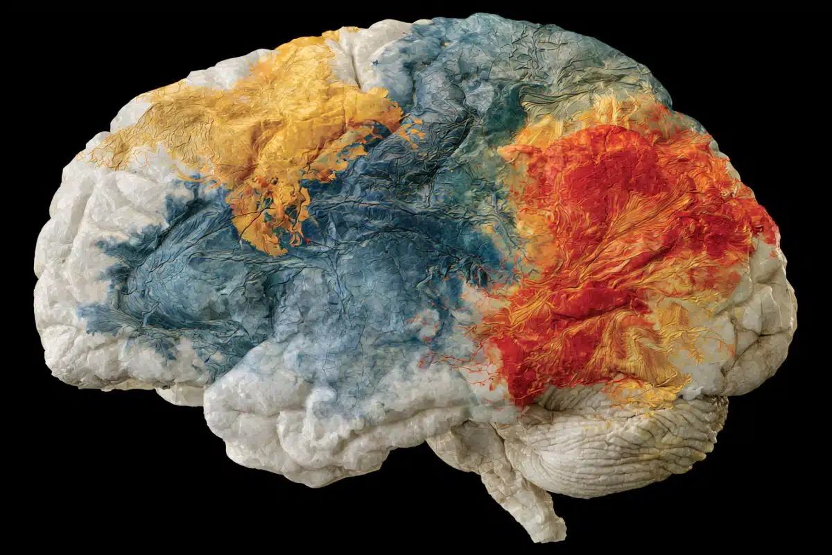
Summary: Researchers evaluated structural MRI scans from 45,900 controls and 2,698 individuals across 9 conditions to map brain aging signatures. Measuring Predictive Age Difference (PAD), the team found that Alzheimer’s disease and mild cognitive impairment showed the highest accelerated brain aging, followed by psychiatric disorders and substance addiction.
ADHD and autism showed no increase in PAD. The study mapped distinct regional aging patterns, such as default mode network involvement in addiction and frontal-temporal acceleration in psychiatric conditions, offering new structural biomarkers for clinical neuroscience.
Key Facts
- Accelerated Aging Rankings: Neurodegenerative conditions (Alzheimer’s disease and MCI) showed the highest overall positive PAD (most pronounced accelerated brain aging), followed by psychiatric disorders and substance addictions.
- Neurodevelopmental Divergence: Attention-deficit/hyperactivity disorder (ADHD) and autism spectrum disorder (ASD) showed no significant increase in PAD compared to healthy controls, indicating that neurodivergence does not equate to accelerated structural brain aging.
- Regional Aging Signatures:
- Prefrontal Cortex: Exhibited elevated PAD broadly across multiple brain disorders.
- Frontal & Temporal Lobes: Showed elevated PAD specifically associated with psychiatric disorders.
- Frontal & Occipital Cortex: Showed localized accelerated aging signature patterns in dementia.
- Default Mode & Salience Networks: Showed selective elevated PAD tied to alcohol and tobacco addiction, alongside structural shifts in the putamen and thalamus.
- Transcriptomic Link: Regional PAD maps correlated with condition-specific gene transcription patterns, offering biological insights into the pathways underlying accelerated structural decline.
Source: PLOS
People with dementia, mild cognitive impairment, alcohol addiction, or psychiatric disorders such as schizophrenia show increased brain aging, each in specific patterns within the brain, according to a study published July 21st in the open access journal PLOS Medicine by Shile Qi from the Nanjing University of Aeronautics and Astronautics, China, and colleagues.
Some conditions can make the brain age faster. Scientists calculate how old the brain is relative to the body using the predictive age difference (PAD), the difference between chronological age and the age predicted by brain imagine, where a positive PAD indicates that aging is accentuated or increased.
To better understand how brain disorders and divergences might affect brain aging, the authors of this study collected structure magnetic resonance imaging (MRI) data from 45,900 controls across several brain imaging banks, and compared them with of 2,698 patients with different brain conditions and differences, including attention-deficit/hyperactivity disorder (ADHD), autism spectrum disorder (ASD), alcohol or tobacco addiction, Alzheimer’s disease (AD), mild cognitive impairment (MCI), schizophrenia, bipolar disorder or major depressive disorder.
The authors found that neurodegenerative disorders of AD and MCI had the largest association with a high PAD. Addiction and psychiatric disorders were also associated with increased PAD. In contrast, there were no differences in PAD between people with ADHD or ASD and controls.
The researchers also looked at PAD values in specific areas of the brain, and examined which genes showed increased expression in people with different brain conditions.
The prefrontal cortex showed higher PAD across brain disorders. Higher PAD in the frontal and temporal lobes was associated with psychiatric disorders, while high PAD in the frontal and occipital cortex was associated with dementia.
Addiction was connected with high PAD in the default mode network, and in the salience network and the putamen and thalamus. There were also differences in gene transcription that associated with specific conditions and divergences.
While the results are correlational, and not causal, and while some conditions such as psychiatric disorders and addiction have high co-occurrence, the author suggest that understanding more about PAD could help provide biomarkers for commonly occurring brain disorders.
The authors add, “Different neurological disorders appear to leave different signatures on the brain aging clock, which may help researchers better understand the neural and biological pathways involved in these conditions.”
Funding: This work was supported by the Key Research and Development Plan of Jiangsu Province, China (BE2023668, https://kxjst.jiangsu.gov.cn) to S.Q., and the National Natural Science Foundation of China (62376124, https://www.nsfc.gov.cn) to S.Q. The funders had no role in study design, data collection and analysis, decision to publish, or preparation of the manuscript.
Key Questions Answered:
A: Predictive Age Difference (PAD) is calculated by comparing a person’s actual chronological age with their estimated “brain age,” derived from structural MRI scans analyzed via machine learning algorithms. A positive PAD value indicates that the structural features of the brain resemble those of a chronologically older individual, signaling accelerated brain aging.
A: No. While neurodegenerative conditions (like Alzheimer’s and MCI), psychiatric disorders (like schizophrenia and major depression), and addictions (alcohol and tobacco) showed increased PAD, neurodevelopmental conditions like ADHD and autism spectrum disorder (ASD) showed no increase in brain aging compared to healthy controls.
A: Broad brain aging metrics only tell part of the story. By mapping accelerated aging to specific circuits, such as the default mode network in addiction or the temporal lobe in psychiatric illness, researchers can identify distinct biological pathways, potential biomarkers for early diagnosis, and targeted circuit interventions.
Editorial Notes:
- This article was edited by a Neuroscience News editor.
- Journal paper reviewed in full.
- Additional context added by our staff.
About this neurology and brain aging research news
Author: Claire Turner
Source: PLOS
Contact: Claire Turner – PLOS
Image: The image is credited to Neuroscience News
Original Research: Open access.
“Brain aging patterns among nine neurological disorders: A case-control study” by Chuang Liang, Godfrey Pearlson, Juan Bustillo, Peter Kochunov, Jiayu Chen, Xiangrong Zhang, Rongtao Jiang, Kent E. Hutchison, Jing Sui, Zening Fu, Xiao Yang, Yuhui Du, Daoqiang Zhang, Shile Qi, Vince D. Calhoun. PLOS Medicine
DOI:10.1371/journal.pmed.1004860
Abstract
Brain aging patterns among nine neurological disorders: A case-control study
Background
The difference between neuroimaging-predicted brain age and chronological age, the predicted age difference (PAD), has been studied as a potential biomarker reflecting individual brain health.
Although previous large-scale studies have shown that brain age deviations occur across multiple disorders, cross-disorder comparisons of PAD within a unified framework, together with identification of the neuroimaging features associated with these differences and their related gene expression profiles, remain limited.
Our aims are to systematically compare brain aging across multiple common brain disorders and explore the brain patterns and biological processes underlying these differences.
Methods and findings
In this study, structural MRI data from 45,900 healthy controls (HCs) and 2,698 patients with developmental disorders (attention-deficit/hyperactivity disorder [ADHD] and autism spectrum disorder [ASD]), addiction (alcohol use disorder [AUD], tobacco use disorder [TUD], and AUD&TUD-A&TUD), dementia (Alzheimer’s disease [AD], and mild cognitive impairment [MCI]) or other psychiatric disorders (schizophrenia [SZ], bipolar disorder [BP], and major depressive disorder [MDD]), were collected to generate PAD, along with transcriptome data.
Then, we calculated the PAD difference between patient and HC as Cohen’s d effect sizes, derived from a linear model that accounted for age, age2, sex, and site, and further identified the interpretable brain patterns associated with the PAD difference for each diagnostic group.
Finally, enrichment analyses was conducted to identify the biological function of genes relatively over- or underexpressed in association with these patterns.
Results showed that while PAD was consistently greater across disorders, different brain disorders showed different degrees of abnormality, the highest effects in dementia (AD: d = 0.97, 95% confidence interval (CI) [0.82,1.13]; p < 0.001 and MCI: d = 0.45, 95% CI [0.34,0.56]; p < 0.001), followed by addiction (A&TUD: d = 0.84, 95% CI [0.44,1.23]; p < 0.001, TUD: d = 0.72, 95% CI [0.49,0.96]; p < 0.001, and AUD d = 0.62, 95% CI [0.39,0.84]; p < 0.001) and psychiatric disorders (SZ: d = 0.53, 95% CI [0.30,0.76]; p < 0.001, BP: d = 0.46, 95% CI [0.22,0.69]; p < 0.001 and MDD: d = 0.28, 95% CI [0.11,0.46]; p < 0.001), but not different from expected in developmental disorders (ASD: d = 0.06, 95% CI [−0.04,0.16]; p = 0.36) and ADHD: d = 0.01, 95% CI [−0.14,0.15]; p = 0.98).
Furthermore, higher PAD values in patient groups were linked to specific spatial brain patterns, including the frontotemporal network in psychiatric disorders, default mode network-salience network-putamen-thalamus in addiction and fronto-occipital network in dementia.
Prefrontal cortex involvement was common across disorders, and disorder-specific brain patterns associated genes were enriched in different biological processes. A limitation of our study is that psychiatric disorders and addiction have high comorbidity, and these potential confounders were not considered.
Conclusions
In summary, the different brain aging patterns, each based around specific underlying circuits, may serve as neuroimaging biomarkers for understanding the neural aging mechanisms in commonly occurring brain disorders. Future studies should test whether these disorder-specific brain aging patterns can serve as useful biomarkers to guide critical clinical decision-making.


