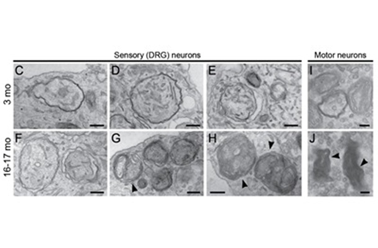
Summary: In aging mice, the number of autophagosomes is significantly decreased, and pronounced defects in the structure of autophagosomes increased. Turning on the WIPI2B protein restores autophagosome formation in aged neurons.
Source: University of Pennsylvania
Cells dispose of harmful “trash” through autophagy, a normal and necessary process in which aggregated proteins and dysfunctional structures are handled. If any part of this fails, waste builds up inside cells, eventually killing them. According to a new study from the Perelman School of Medicine at the University of Pennsylvania, as cells age, their ability to shed harmful refuse declines. The findings suggest that the deterioration of autophagy in aged neurons—cells that never replicate and are as old as the bodies they inhabit—could be a risk factor for a suite of neurodegenerative diseases such as ALS (amyotrophic lateral sclerosis), Alzheimer’s, and Parkinson’s.
Using live-cell imaging of neurons from young and aged mice, Erika Holzbaur, Ph.D., a professor of Physiology, and first author Andrea Stavoe, Ph.D., a postdoctoral fellow in Holzbaur’s lab, published their study this week in eLife. The importance of autophagy was recognized in 2016 with the Nobel Prize in Physiology or Medicine.
“The current thinking among scientists is that a decline in autophagy makes neurons more vulnerable to genetic or environmental risks,” Holzbaur said. “What motivates our line of research is that most neurodegenerative diseases in which a deterioration of autophagy has been implicated, such as ALS, and Alzheimer’s, Huntington’s and Parkinson’s diseases, are also disorders of aging,”
At the start of autophagy, a component within the cell called an autophagosome, engulfs misfolded proteins or damaged structures to be degraded, essentially sequestering this waste in a biological trash bag. The autophagosome then fuses with a second cellular structure, called a lysosome, that contains the enzymes needed to breakdown the garbage, allowing the components to be recycled and reused. This elegant waste-removal stream is what keeps neurons healthy, but in its absence, neurons eventually die due to the buildup of unattended refuse.
“Think city streets during a sanitation workers strike,” Stavoe said.
The team assessed rates of autophagy in mouse neurons during aging and identified a significant decrease in the number of autophagosomes produced, along with pronounced defects in the structure of autophagosomes produced by neurons from aged mice.
While early stages of autophagosome formation were unaffected, they found frequent stalling in their formation in aged mice, while the ones that did form were misshapen. These defects may allow the trash to accumulate at neuronal synapses. Stavoe notes that in other studies autophagosomes with misformed membranes have been observed in deceased human brain tissue from donors with neurodegenerative disease.
Importantly, turning on the protein WIPI2B in aged mice restores autophagosome formation in aged neurons, bringing the autophagy garbage-hauling process back online. This rescue is dependent on the level of activation of WIPI2B, providing insight into the biological regulation of autophagosome formation.
On the other hand, when researchers took WIPI2B out of young neurons, autophagosome formation stalled. “This stunning and complete rescue of autophagy using one protein suggests a novel therapeutic target for age-associated neurodegeneration,” Stavoe said.
Other co-authors are Pallavi Gopal from Yale University, and Andrea Gubas and Sharon Tooze, from The Francis Crick Institute, London.
Funding: This work was funded with an NIH Pathways to Independence Fellowship (K99 NS109286) and a Javits Award from NINDS (R37 NS060698).
Source:
University of Pennsylvania
Media Contacts:
Karen Kreeger – University of Pennsylvania
Image Source:
The image is credited to Andrea Stavoe, Penn Medicine; eLife.
Original Research: Open access
“Expression of WIPI2B counteracts age-related decline in autophagosome biogenesis in neurons”. Andrea KH Stavoe, Pallavi P Gopal, Andrea Gubas, Sharon A Tooze, Erika LF Holzbaur.
eLife. doi:10.7554/eLife.44219
Abstract
Expression of WIPI2B counteracts age-related decline in autophagosome biogenesis in neurons
Autophagy defects are implicated in multiple late-onset neurodegenerative diseases including Amyotrophic Lateral Sclerosis (ALS) and Alzheimer’s, Huntington’s, and Parkinson’s diseases. Since aging is the most common shared risk factor in neurodegeneration, we assessed rates of autophagy in mammalian neurons during aging. We identified a significant decrease in the rate of constitutive autophagosome biogenesis during aging and observed pronounced morphological defects in autophagosomes in neurons from aged mice. While early stages of autophagosome formation were unaffected, we detected the frequent production of stalled LC3B-negative isolation membranes in neurons from aged mice. These stalled structures recruited the majority of the autophagy machinery, but failed to develop into LC3B-positive autophagosomes. Importantly, ectopically expressing WIPI2B effectively restored autophagosome biogenesis in aged neurons. This rescue is dependent on the phosphorylation state of WIPI2B at the isolation membrane, suggesting a novel therapeutic target in age-associated neurodegeneration.