

Summary: Researchers pioneered a new method to monitor blood vessel dynamics in the mouse brain, revealing that visual stimuli can synchronize vasomotion, potentially improving brain function. By exposing mice to a specific pattern of horizontally moving stripes, the team observed that vasomotion matched the stimulus pattern’s speed and spread throughout the brain.
This synchronization suggests a mechanism by which the brain enhances its circulation of nutrients and clears waste, possibly boosting cognitive abilities. The findings could have implications for treating and preventing neurological conditions.
Key Facts:
- Innovative Monitoring Technique: The study introduces a novel method to observe blood vessel dynamics through the intact skull or deep within the brain using optical fibers.
- Vasomotion Synchronization: Visual stimuli can entrain the brain’s vasomotion, leading to synchronized blood vessel activity that enhances nutrient delivery and waste removal across the entire brain.
- Potential Health Benefits: This synchronized vasomotion may improve learning, aid in stroke recovery, and potentially delay neurodegenerative diseases like dementia by improving the brain’s metabolic efficiency.
Source: Tohoku University
Compared with computers, the brain can perform computations with a very low net energy supply. Yet our understanding surrounding how the biological brain manages energy is still incomplete.
What is known, however, is that the dilation and constriction cycles of blood vessels, or vasomotion, spontaneously occur in the brain, a process that likely contributes to enhancing the circulation of energetic nutrients and clearing wasteful materials.
Now, researchers from Tohoku University have developed a method that easily observes and monitors blood vessel dynamics in the mouse brain. This can be done either through the intact skull of a mouse, or deep into the brain using an implanted optical fiber.
The findings were detailed in the journal, eLife, on April 17, 2024.
Since it has been reported that sensory stimuli can cause dilation of blood vessels or hyperemia, researchers sought to induce vasomotion via presenting mice with visual stimuli.
What they discovered was when a mouse was shown a horizontally moving stripe pattern that changed direction every 2 to 3 seconds, it caused a reaction in the mouse’s blood vessels that matched the pattern’s speed.
Mice were presented with 15-minute visual training sessions interleaved with 1-hour resting periods for 4 times per day. With such spaced training, the amplitude of the synchronized vasomotion gradually increased.
Interestingly, the visually induced vasomotion was not confined to the area of the cerebral cortex responsible for visual information processing. In other words, synchronized vasomotion spread throughout the whole brain.
“Synchronized vascular motion can be entrained with slowly oscillating visual stimuli,” says Professor Ko Matsui of the Super-network Brain Physiology lab at Tohoku University, who led the research.
“Such enhancement of circulation mechanisms may benefit the information processing capacity of the brain.”
While it’s long been known that changes in neuron connections support learning and memory, the plasticity of vasomotion hasn’t been described before.
Matsui and his colleagues found that a specific visual pattern makes the eyes move more, and this eye movement improvement depends on changes in the brain’s cerebellum. The researchers also observed that blood vessel activity in the cerebellum synchronized with this optokinetic motor learning.
Lead study investigator, Daichi Sasaki, believes that synchronized vasomotion, which efficiently delivers oxygen and glucose, could improve learning abilities.
He states, “Our next step is to explore the advantages of vasomotion synchronization. It might help clear waste like amyloid beta, potentially delaying or preventing dementia.
“Stroke recovery could also benefit from better energy supply and waste removal. Additionally, synchronized vasomotion might even enhance intelligence beyond our natural capabilities.”
About this cognition and neuroscience research news
Author: Public Relations
Source: Tohoku University
Contact: Public Relations – Tohoku University
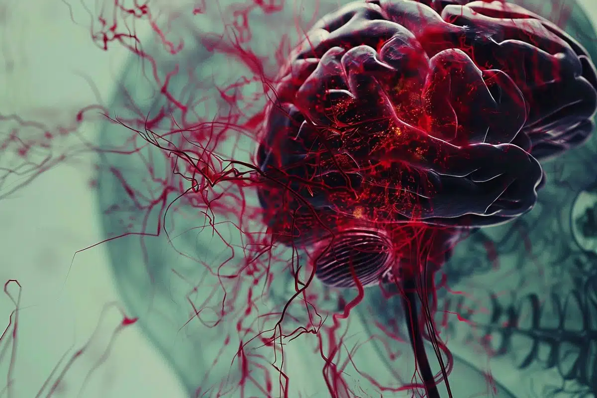
Image: The image is credited to Neuroscience News
Original Research: Open access.
“Plastic vasomotion entrainment” by Daichi Sasaki et al. eLife
Abstract
Plastic vasomotion entrainment
The presence of global synchronization of vasomotion induced by oscillating visual stimuli was identified in the mouse brain.
Endogenous autofluorescence was used and the vessel ‘shadow’ was quantified to evaluate the magnitude of the frequency-locked vasomotion.
This method allows vasomotion to be easily quantified in non-transgenic wild-type mice using either the wide-field macro-zoom microscopy or the deep-brain fiber photometry methods.
Vertical stripes horizontally oscillating at a low temporal frequency (0.25 Hz) were presented to the awake mouse, and oscillatory vasomotion locked to the temporal frequency of the visual stimulation was induced not only in the primary visual cortex but across a wide surface area of the cortex and the cerebellum.
The visually induced vasomotion adapted to a wide range of stimulation parameters. Repeated trials of the visual stimulus presentations resulted in the plastic entrainment of vasomotion.
Horizontally oscillating visual stimulus is known to induce horizontal optokinetic response (HOKR). The amplitude of the eye movement is known to increase with repeated training sessions, and the flocculus region of the cerebellum is known to be essential for this learning to occur.
Here, we show a strong correlation between the average HOKR performance gain and the vasomotion entrainment magnitude in the cerebellar flocculus. Therefore, the plasticity of vasomotion and neuronal circuits appeared to occur in parallel.
Efficient energy delivery by the entrained vasomotion may contribute to meeting the energy demand for increased coordinated neuronal activity and the subsequent neuronal circuit reorganization.


