Summary: Researchers have shown that astrocytes in the lateral hypothalamus play a pivotal role in how stress shapes behavior, particularly after early-life adversity. In mice, stress led to smaller, less branched astrocytes and abnormal orexin neuron activity, with sex-specific patterns in activity levels.
By deleting stress hormone receptors in astrocytes, the scientists were able to restore normal behavior and neuronal function, even if astrocytes didn’t fully return to their original size. The findings highlight astrocytes as potential therapeutic targets for preventing treatment-resistant depression in humans.
Key Facts
- Astrocyte Role: Stress disrupts astrocyte structure and function before neurons are affected.
- Sex Differences: Early-life stress caused opposite behavioral changes in male and female mice.
- Therapeutic Potential: Removing stress hormone receptors in astrocytes normalized behavior and neuronal activity.
Source: University of Montreal
Astrocytes in the lateral hypothalamus region of the brain, an area involved in the regulation of sleep and wakefulness, play a key role in neuron activity in mice and affect their behaviour, Canadian researchers have found.
Led by Ciaran Murphy-Royal of Université de Montréal’s affiliated hospital research centre, the CRCHUM, the scientists detail their finding in a study published in Nature Communications.

In so broadening medical science’s understanding of cerebral mechanisms, the discovery could someday help in the treatment and prevention of depression in humans, the researchers say.
According to the scientific literature, early-life stress leads to a five-fold increase in the risk of developing a mental-health disorder as an adult, notably causing treatment-resistant disorders.
Less active, day or night
As brain cells, astrocytes are sensitive to variations in the blood concentration of metabolites and, in response to changes in the blood, astrocytes can modulate the extent of their interaction with neurons, their neighbouring cells.
In mice, those changes are particularly responsive to the level of corticosterone, the stress hormone in the rodents’ blood.
“In adult mice who experienced early-life stress, we saw abnormally high levels of corticosterone,” said Murphy-Royal, a professor in UdeM’s Faculty of Medicine. “The impact of stress on behaviour also differed according to sex.”
Notably, he said, “females were less active at night, while males were hyperactive during the day.”
In people with depression who have experienced a similar type of stress, this sex differences have also been observed.
Lack of maternal care
Lewis R. Depaauw-Holt, the study’s first author and a PhD student on Murphy-Royal’s team, was able to recreate early-life stress conditions in young rodents by separating them from their mothers.
Over 10 days, for four hours a day, he kept the young mice apart from their mothers. This lack of maternal care occurred during a critical period of brain development for the rodents, the equivalent of ages three to seven in human children.
“The differences in activity levels between female and male mice were also seen within a group of neurons that produces neuropeptides called orexins,” said Murphy-Royal.
“Located in the lateral hypothalamus, these orexin neurons contribute to regulating sleep-wake cycles,” he said. “In males, these neurons showed hyperactivity, while in females we saw hypoactivity.”
In mice that experienced early-life stress, astrocytes were smaller and had fewer branches, especially in females. These branches are essential for transmitting information to neighbouring neurons and interacting with nearby cells.
“In our field of expertise, we believe that the changes in astrocyte morphology are a marker of dysfunction,” said Murphy-Royal. “In humans, we see these variations in diseases like Parkinson’s or Alzheimer’s.”
A single pathway?
What if these changes in behaviour, neuron activity and morphology in both sexes were tied to a single stress-signalling pathway?
To test their hypothesis, the CRCHUM research team deleted glucocorticoid receptors in astrocytes to which corticosterone, the stress hormone, would normally bind.
“Without these receptors, neuronal activity and mouse behaviours returned to baseline similar to that of mice who did not experience early-life stress,” said Murphy-Royal.
“And, even if their astrocytes didn’t return to their normal size, they did regain their complexity, visible in the number of branches they use to interact with neighbouring cells.”
Contrary to what scientists believed until now, astrocytes are disturbed by stress before neurons are, the study reveals.
In humans, the challenge of countering the effects of early-life stress will surely be more complex than in rodents, Murphy-Royal cautioned, but one thing is for sure: astrocytes could turn out to be an excellent therapeutic target for preventing depression.
About this neuroscience and mental health research news
Author: Bruno Geoffroy
Source: University of Montreal
Contact: Bruno Geoffroy – University of Montreal
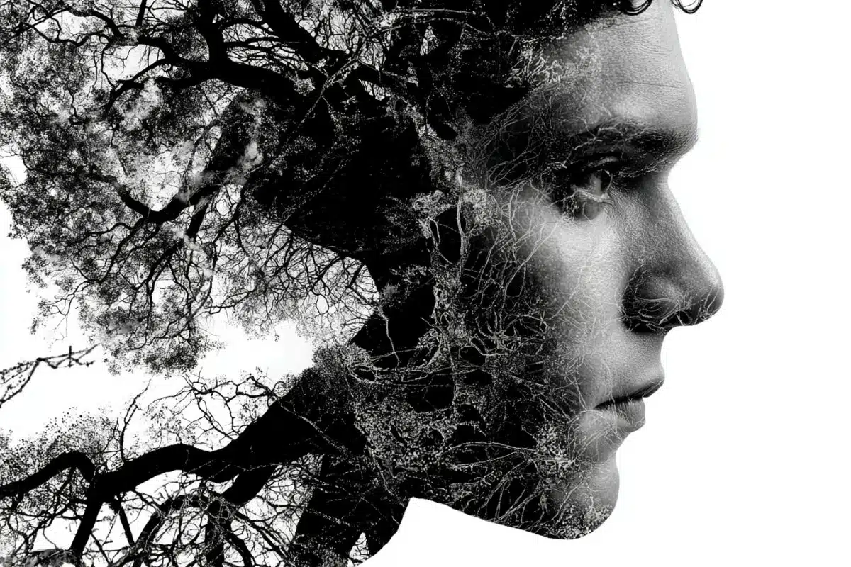
Image: The image is credited to Neuroscience News
Original Research: Open access.
“A divergent astrocytic response to stress alters activity patterns via distinct mechanisms in male and female mice” by Ciaran Murphy-Royal et al. Nature Communications
Abstract
A divergent astrocytic response to stress alters activity patterns via distinct mechanisms in male and female mice
The lateral hypothalamus is a brain region that regulates activity levels, circadian, and motivated behaviour.
While disruption of these behaviours forms a hallmark of stress-related neuropsychiatric disorders, the underlying cellular mechanisms of how stress affects this brain region remain poorly understood.
Here, we report that the effects of stress on behavioural activity levels correlate with spontaneous firing of orexin neurons, inducing hyperactivity in males and hypoactivity in female mice.
These neuronal changes are accompanied by astrocyte remodelling, with causal manipulations identifying lateral hypothalamic astrocytes as key regulators of neuronal firing and physical activity patterns.
In the context of stress, sex-specific changes in orexin neuron firing were driven by distinct astrocytic mechanisms with elevated purinergic signaling in male mice and reduced extracellular L-lactate in female mice.
Finally, we show that genetic deletion of glucocorticoid receptors in lateral hypothalamic astrocytes restores key aspects of astrocyte morphology, rescues the effects of stress on orexin neuron firing, and recovers activity levels in both males and females.
Overall, these data causally implicate astrocytes in the regulation of orexin neuron firing, behavioural activity patterns, and reveal that astrocytes are primary drivers of stress-induced behavioural change.