

A method for detecting early signs of Alzheimer’s disease using amyloid PET imaging works as well as the previously used cerebrospinal fluid sample method. This is the conclusion of a new Lund University study – the most thorough and extensive undertaken in the field so far.
The most commonly used tools for investigating early signs of Alzheimer’s disease in Swedish public healthcare are various cognitive memory tests and computed tomography. For several years it has also been possible to carry out an analysis of a cerebrospinal fluid sample which increases the chances of early detection. So far, however, only patients in memory clinics have been offered the test.
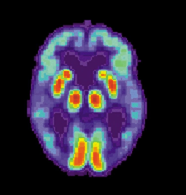
Recently, a method known as amyloid PET was approved for clinical use in Sweden. A special substance which binds to a protein in the brain, β-amyloid, is administered to the patient. This amyloid is a marker for Alzheimer’s changes, which are then mapped with PET imaging.
Opinions have long been divided as to whether cerebrospinal fluid samples or PET imaging are the best tools for detecting early-stage Alzheimer’s disease.
“In the study, both the cerebrospinal fluid sample and the amyloid PET scans were able to identify approximately 90 per cent of the patients who would be diagnosed with Alzheimer’s later on. Our conclusion is therefore that the two methods work equally well to achieve this aim. One can thus choose the method on the basis of cost, expertise or patient preference”, says Sebastian Palmqvist, MD, PhD, at Lund University.
Both methods are also good at identifying which individuals are healthy and unlikely to develop Alzheimer’s disease within the next ten years. However, when the diagnosis is reached without reference to a cerebrospinal fluid sample or amyloid PET imaging, its accuracy can drop to 60-70 per cent.
Late detection of Alzheimer’s disease is not only a problem for today’s healthcare, but also for the development of future treatments.
“Previous drug trials to evaluate new treatments for the presence of amyloid in Alzheimer’s cases failed, partly because treatment began too late in the course of the disease. With two accurate tools for early diagnosis, we can identify suitable participants at an early stage of Alzheimer’s disease. This will considerably increase the chances of being able to prove a positive effect for new drugs”, concludes Oskar Hansson, associate professor and neurologist at Lund University.
The research data originates from the Swedish BioFINDER study. 122 healthy elderly participants and 34 patients with mild cognitive impairment who developed Alzheimer’s disease within three years were investigated in the article. The study was then repeated in an American population group of 210 individuals. The new findings are presented in the American journal Neurology.
Funding: Funders of the study include the Swedish Research Council, the European Research Council, Region Skåne and MultiPark at Lund University.
Source: Sebastian Palmqvist – Lund University
Image Credit: The image is credited to the NIH and is in the public domain
Original Research: Abstract for “Detailed comparison of amyloid PET and CSF biomarkers for identifying early Alzheimer disease” by Sebastian Palmqvist, MD, PhD, Henrik Zetterberg, MD, PhD, Niklas Mattsson, MD, PhD, Per Johansson, MD, PhD; For the Alzheimer’s Disease Neuroimaging Initiative, Lennart Minthon, MD, PhD, Kaj Blennow, MD, PhD, Mattias Olsson, PhD; For the Swedish BioFINDER study group and Oskar Hansson, MD, PhD in Neuron. Published online September 9 2015 doi:10.1212/WNL.0000000000001991
Abstract
Detailed comparison of amyloid PET and CSF biomarkers for identifying early Alzheimer disease
Objective: To compare the diagnostic accuracy of CSF biomarkers and amyloid PET for diagnosing early-stage Alzheimer disease (AD).
Methods: From the prospective, longitudinal BioFINDER study, we included 122 healthy elderly and 34 patients with mild cognitive impairment who developed AD dementia within 3 years (MCI-AD). β-Amyloid (Aβ) deposition in 9 brain regions was examined with [18F]-flutemetamol PET. CSF was analyzed with INNOTEST and EUROIMMUN ELISAs. The results were replicated in 146 controls and 64 patients with MCI-AD from the Alzheimer’s Disease Neuroimaging Initiative study.
Results: The best CSF measures for identifying MCI-AD were Aβ42/total tau (t-tau) and Aβ42/hyperphosphorylated tau (p-tau) (area under the curve [AUC] 0.93–0.94). The best PET measures performed similarly (AUC 0.92–0.93; anterior cingulate, posterior cingulate/precuneus, and global neocortical uptake). CSF Aβ42/t-tau and Aβ42/p-tau performed better than CSF Aβ42 and Aβ42/40 (AUC difference 0.03–0.12, p < 0.05). Using nonoptimized cutoffs, CSF Aβ42/t-tau had the highest accuracy of all CSF/PET biomarkers (sensitivity 97%, specificity 83%). The combination of CSF and PET was not better than using either biomarker separately.
Conclusions: Amyloid PET and CSF biomarkers can identify early AD with high accuracy. There were no differences between the best CSF and PET measures and no improvement when combining them. Regional PET measures were not better than assessing the global Aβ deposition. The results were replicated in an independent cohort using another CSF assay and PET tracer. The choice between CSF and amyloid PET biomarkers for identifying early AD can be based on availability, costs, and doctor/patient preferences since both have equally high diagnostic accuracy.
Classification of evidence: This study provides Class III evidence that amyloid PET and CSF biomarkers identify early-stage AD equally accurately.
“Detailed comparison of amyloid PET and CSF biomarkers for identifying early Alzheimer disease” by Sebastian Palmqvist, MD, PhD, Henrik Zetterberg, MD, PhD, Niklas Mattsson, MD, PhD, Per Johansson, MD, PhD; For the Alzheimer’s Disease Neuroimaging Initiative, Lennart Minthon, MD, PhD, Kaj Blennow, MD, PhD, Mattias Olsson, PhD; For the Swedish BioFINDER study group and Oskar Hansson, MD, PhD in Neuron. Published online September 9 2015 doi:10.1212/WNL.0000000000001991


