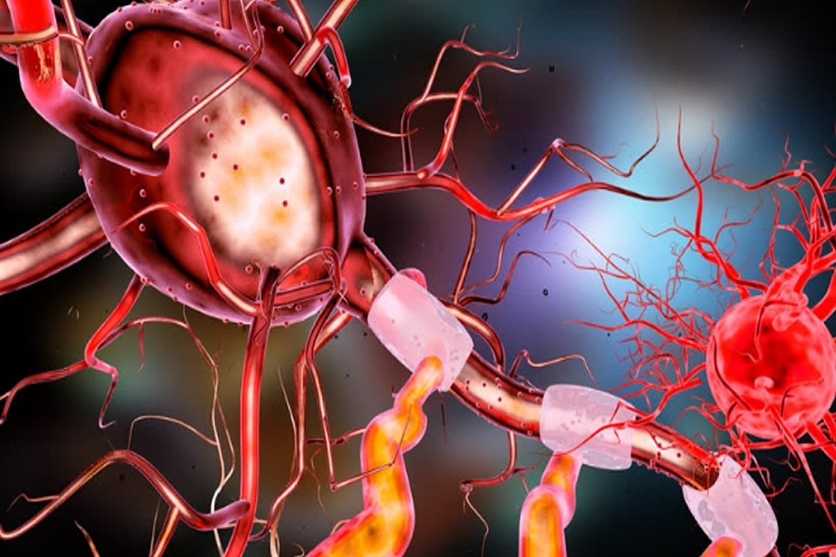
Summary: Microglia may play a significant role in disrupting neurogenesis in those with Alzheimer’s disease. When mice were given drugs which caused microglial cells to die, neurogenesis returned to normal.
Source: University of Chicago
Much of the research on the underlying causes of Alzheimer’s disease focuses on amyloid beta (Aß), a protein that accumulates in the brain as the disease progresses. Excess Aß proteins form clumps or “plaques” that disrupt communication between brain cells and trigger inflammation, eventually leading to widespread loss of neurons and brain tissue.
Aß plaques will continue to be a major focus for Alzheimer’s researchers. However, new work by neuroscientists at the University of Chicago looks at another process that plays an underappreciated role in the progression of the disease.
In a new study published in the Journal of Neuroscience, Sangram Sisodia, PhD, the Thomas Reynolds Sr. Family Professor of Neurosciences at UChicago, and his colleagues show how in genetic forms of Alzheimer’s, a process called neurogenesis, or the creation of new brain cells, can be disrupted by the brain’s own immune cells.
Some types of early onset, hereditary Alzheimer’s are caused by mutations in two genes called presenilin 1 (PS1) and presenilin 2 (PS2). Previous research has shown that when healthy mice are placed into an “enriched” environment where they can exercise, play and interact with each other, they have a huge increase in new brain cells being created in the hippocampus, part of the brain that is important for memory. But when mice carrying mutations to PS1 and PS2 are placed in an enriched environment, they don’t show the same increase in new brain cells. They also start to show signs of anxiety, a symptom often reported by people with early onset Alzheimer’s.
This led Sisodia to think that something besides genetics had a role to play. He suspected that the process of neurogenesis in mice both with and without Alzheimer’s mutations could also be influenced by other cells that interact with the newly forming brain cells.
Focus on the microglia
The researchers focused on microglia, a kind of immune cell in the brain that usually repairs synapses, destroys dying cells and clears out excess Aß proteins. When the researchers gave the mice a drug that causes microglial cells to die, neurogenesis returned to normal. The mice with presenilin mutations were then placed into an enriched environment and they were fine; they didn’t show any memory deficits or signs of anxiety, and they were creating the normal, expected number of new neurons.

“It’s the most astounding result to me,” Sisodia said. “Once you wipe out the microglia, all these deficits that you see in these mice with the mutations are completely restored. You get rid of one cell type, and everything is back to normal.”
Sisodia thinks the microglia could be overplaying their immune system role in this case. Alzheimer’s disease normally causes inflammation in the microglia, so when they encounter newly formed brain cells with presenilin mutations they may overreact and kill them off prematurely. He feels that this discovery about the microglia’s role opens another important avenue toward understanding the biology of Alzheimer’s disease.
“I’ve been studying amyloid for 30 years, but there’s something else going on here, and the role of neurogenesis is really underappreciated,” he said. “This is another way to understand the biology of these genes that we know significantly affect the progression of disease and loss of memory.”
Additional authors include Sylvia Ortega-Martinez, Nisha Palla, Xiaoqiong Zhang and Erin Lipman from the University of Chicago.
Source:
University of Chicago
Media Contacts:
Matt Wood – University of Chicago
Image Source:
The image is in the public domain.
Original Research: Closed access
“Deficits in Enrichment-Dependent Neurogenesis and Enhanced Anxiety Behaviors Mediated by Expression of Alzheimer’s Disease-Linked Ps1 Variants Are Rescued by Microglial Depletion”. Sylvia Ortega-Martinez, Nisha Palla, Xiaoqiong Zhang, Erin Lipman and Sangram S. Sisodia.
Journal of Neuroscience. doi:10.1523/JNEUROSCI.0884-19.2019
Abstract
Deficits in Enrichment-Dependent Neurogenesis and Enhanced Anxiety Behaviors Mediated by Expression of Alzheimer’s Disease-Linked Ps1 Variants Are Rescued by Microglial Depletion
Alzheimer’s disease (AD) is a progressive neurodegenerative disorder that presently affects an estimated 5.7 million Americans. Understanding the basis for this disease is key for the development of a future successful treatment. In this effort, we previously reported that mouse prion protein-promoter-driven, ubiquitous expression of familial AD (FAD)-linked human PSEN1 variants in transgenic mice impairs environmental enrichment (EE)-induced proliferation and neurogenesis of adult hippocampal neural progenitor cells (AHNPCs) and in a non-cell autonomous manner. These findings were confirmed in PS1M146V/+ mice that harbor an FAD-linked mutation in the endogenous PSEN1 gene. We now demonstrate that CSF1R antagonist-mediated microglial depletion in transgenic male mice expressing mutant presenilin 1 (PS1) or PS1M146V/+ “knock-in” mice leads to a complete rescue of deficits in proliferation, differentiation and survival of AHNPCs. Moreover, microglia depletion suppressed the heightened baseline anxiety behavior observed in transgenic mice expressing mutant PS1 and PS1M146V/+ mice to levels observed in mice expressing wild-type human PS1 or nontransgenic mice, respectively. These findings demonstrate that in mice expressing FAD-linked PS1, microglia play a critical role in the regulation of EE-dependent AHNPC proliferation and neurogenesis and the modulation of affective behaviors.
SIGNIFICANCE STATEMENT
Inheritance of mutations in genes encoding presenilin 1 (PS1) causes familial Alzheimer’s disease (FAD). Mutant PS1 expression enhances the levels and assembly of toxic Aβ42 peptides and impairs the self-renewal and neuronal differentiation of adult hippocampal neural progenitor cells (AHNPCs) following environmental enrichment (EE) that is associated with heightened baseline anxiety. We now show that microglial depletion fully restores the EE-mediated impairments in AHNPC phenotypes and suppresses the heightened baseline anxiety observed in mice expressing FAD-linked PS1. Thus, we conclude that the memory deficits and anxiety-related behaviors in patients with PS1 mutations is a reflection not just of an increase in the levels of Aβ42 peptides, but to impairments in the self-renewal and neuronal differentiation of AHNPCs that modulate affective behaviors.


