

Summary: Researchers report groups of brain regions that synchronize their activity during memory tasks become smaller and more numerous as people age.
Source: PLOS.
Groups of brain regions with coordinated activity are consistent for individuals, but shrink with age.
Groups of brain regions that synchronize their activity during memory tasks become smaller and more numerous as people age, according to a study published in PLOS Computational Biology.
Typically, research on brain activity relies on average brain measurements across entire groups of people. In a new study, Elizabeth Davison of Princeton University, New Jersey, and colleagues describe a novel method to characterize and compare the brain dynamics of individual people.
The researchers used functional magnetic resonance imaging (fMRI) to record healthy people’s brain activity during memory tasks, attention tasks, and at rest. For each person, fMRI data was recast as a network composed of brain regions and the connections between them. The scientists then use this network to measure how closely different groups of connections changed together over time.
They found that, regardless of whether a person is using memory, directing attention, or resting, the number of synchronous groups of connections within one brain is consistent for that person. However, between people, these numbers vary dramatically.
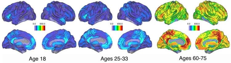
During memory specifically, variations between people are closely linked to age. Younger participants have only a few large synchronous groups that link nearly the entire brain in coordinated activity, while older participants show progressively more and smaller groups of connections, indicating loss of cohesive brain activity–even in the absence of memory impairment.
“This method elegantly captures important differences between individual brains, which are often complex and difficult to describe,” Davison says. “The resulting tools show promise for understanding how different brain characteristics are related to behavior, health, and disease.”
Future work will investigate how to use individual brain signatures to differentiate between healthily aging brains and brains with age-related impairments.
Funding: This work was supported by the David and Lucile Packard Foundation and the Institute for Collaborative Biotechnologies through grant W911NF-09-0001 from the U.S. Army Research Office. KJS was supported by the National Science Foundation Graduate Research Fellowship Program under Grant No. DGE-1144085. END was supported by the National Science Foundation Graduate Research Fellowship Program under Grant No. DGE-1656466 and the Francis Robbins Upton Fellowship in Engineering. END and KJS were additionally supported by the Worster Fellowship. DSB acknowledges support from the John D. and Catherine T. MacArthur Foundation, the Army Research Laboratory and the Army Research Office through contract numbers W911NF-10-2-0022 and W911NF-14-1-0679, the National Institute of Mental Health (2-R01-DC- 009209-11), the National Institute of Child Health and Human Development (1R01HD086888-01), the Office of Naval Research, and the National Science Foundation (#BCS-1441502, #BCS-1430087, and #PHY-1554488). The content of the information does not necessarily reflect the position or the policy of the Government, and no official endorsement should be inferred. The funders had no role in study design, data collection and analysis, decision to publish, or preparation of the manuscript.
Competing Interests: The authors have declared that no competing interests exist.
Source: Elizabeth N. Davison – PLOS
Image Source: NeuroscienceNews.com image is credited to Davison et al.
Original Research: Full open access research for “Individual Differences in Dynamic Functional Brain Connectivity across the Human Lifespan” by Elizabeth N. Davison, Benjamin O. Turner, Kimberly J. Schlesinger, Michael B. Miller, Scott T. Grafton, Danielle S. Bassett, and Jean M. Carlson in PLOS Computational Biology. Published online November 23 2016 doi:10.1371/journal.pcbi.1005178
[cbtabs][cbtab title=”MLA”]PLOS. “Missed Connections: Memory Related Brain Activity Loses Cohesion As We Age.” NeuroscienceNews. NeuroscienceNews, 23 November 2016.
<https://neurosciencenews.com/age-memory-activity-5591/>.[/cbtab][cbtab title=”APA”]PLOS. (2016, November 23). Missed Connections: Memory Related Brain Activity Loses Cohesion As We Age. NeuroscienceNews. Retrieved November 23, 2016 from https://neurosciencenews.com/age-memory-activity-5591/[/cbtab][cbtab title=”Chicago”]PLOS. “Missed Connections: Memory Related Brain Activity Loses Cohesion As We Age.” https://neurosciencenews.com/age-memory-activity-5591/ (accessed November 23, 2016).[/cbtab][/cbtabs]
Abstract
Individual Differences in Dynamic Functional Brain Connectivity across the Human Lifespan
Individual differences in brain functional networks may be related to complex personal identifiers, including health, age, and ability. Dynamic network theory has been used to identify properties of dynamic brain function from fMRI data, but the majority of analyses and findings remain at the level of the group. Here, we apply hypergraph analysis, a method from dynamic network theory, to quantify individual differences in brain functional dynamics. Using a summary metric derived from the hypergraph formalism—hypergraph cardinality—we investigate individual variations in two separate, complementary data sets. The first data set (“multi-task”) consists of 77 individuals engaging in four consecutive cognitive tasks. We observe that hypergraph cardinality exhibits variation across individuals while remaining consistent within individuals between tasks; moreover, the analysis of one of the memory tasks revealed a marginally significant correspondence between hypergraph cardinality and age. This finding motivated a similar analysis of the second data set (“age-memory”), in which 95 individuals, aged 18–75, performed a memory task with a similar structure to the multi-task memory task. With the increased age range in the age-memory data set, the correlation between hypergraph cardinality and age correspondence becomes significant. We discuss these results in the context of the well-known finding linking age with network structure, and suggest that hypergraph analysis should serve as a useful tool in furthering our understanding of the dynamic network structure of the brain.
“Individual Differences in Dynamic Functional Brain Connectivity across the Human Lifespan” by Elizabeth N. Davison, Benjamin O. Turner, Kimberly J. Schlesinger, Michael B. Miller, Scott T. Grafton, Danielle S. Bassett, and Jean M. Carlson in PLOS Computational Biology. Published online November 23 2016 doi:10.1371/journal.pcbi.1005178


