

Summary: Researchers have identified how our brains are so good at perceiving contours and edges. The study, published in Nature, reports neurons are most likely to connect if they react to edges that lie on a common axis and the structure of the world around us is mirrored in the pattern of synapses.
Source: University of Basel.
The research team of Prof. Sonja Hofer at the Biozentrum, University of Basel, has discovered why our brain might be so good at perceiving edges and contours. Neurons that respond to different parts of elongated edges are connected and thus exchange information. This can make it easier for the brain to identify contours of objects. The results of the study are now published in the journal “Nature”.
Individual visual stimuli are not processed independently by our brain. Rather neurons exchange incoming information to form a coherent perceptual image from the myriad of visual details impinging on our eyes. How our visual perception arises from these interactions is still unclear. This is partly due to the fact that we still know relatively little about the rules that determine which neurons in the brain are connected to each other, and what information they exchange. The research team of Prof. Sonja Hofer at the Biozentrum, University Basel studies neuronal networks in the brain. She has now investigated in the mouse model what information individual neurons in the visual cortex receive from other neurons about the wider visual field.
Neurons receive information from large parts of the visual field
The visual cortex, the largest part of the human brain, is responsible for analyzing information from the eyes and enables us to perceive the visual world. Different neurons in this brain area react to components of the visual scene at specific positions in our visual field. Sonja Hofer and her team could show that individual neurons also receive extensive additional information from the remaining visual field. “This is not surprising, because how we perceive individual visual stimuli strongly depends on their surrounding visual environment”, Hofer explains. Individual parts of an image are, for instance, merged into lines, contours and objects.
Edges in our environment are mirrored in the brain
The new study shows that neurons are most likely to be connected if they react to edges that lie on a common axis. “Our visual environment contains many long lines and contours”, Sonja Hofer explains. “The structure of the world around us is therefore mirrored in the pattern of synapses in the brain”. Hofer’s team believes that this specific brain connectivity might facilitate the perception of elongated lines and edges: neurons that react to different parts of such edges are connected, can increase each other’s activity and therefore boost the response that contributes to the perception of these visual features.
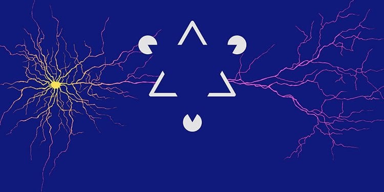
Our brain is so good at identifying contours and objects in images that it is sometimes deceived into seeing them even if they do not actually exist (such as the edges of the blue triangle in the foreground of the figure). Such optical illusions show how primed our brain is to detect lines and object contours”, says Hofer. “Our findings reveal a mechanism that can contribute to this skill”.
Source: Sonja B. Hofer – University of Basel
Image Source: NeuroscienceNews.com image is credited to the researchers.
Original Research: Abstract for “Synaptic organization of visual space in primary visual cortex” by M. Florencia Iacaruso, Ioana T. Gasler & Sonja B. Hofer in Nature. Published online July 12 2017 doi:10.1038/nature23019
[cbtabs][cbtab title=”MLA”]University of Basel “Synapses in the Brain Mirror the Structure of the Visual World.” NeuroscienceNews. NeuroscienceNews, 13 July 2017.
<https://neurosciencenews.com/visual-world-synapses-7073/>.[/cbtab][cbtab title=”APA”]University of Basel (2017, July 13). Synapses in the Brain Mirror the Structure of the Visual World. NeuroscienceNew. Retrieved July 13, 2017 from https://neurosciencenews.com/visual-world-synapses-7073/[/cbtab][cbtab title=”Chicago”]University of Basel “Synapses in the Brain Mirror the Structure of the Visual World.” https://neurosciencenews.com/visual-world-synapses-7073/ (accessed July 13, 2017).[/cbtab][/cbtabs]
Abstract
Synaptic organization of visual space in primary visual cortex
How a sensory stimulus is processed and perceived depends on the surrounding sensory scene. In the visual cortex, contextual signals can be conveyed by an extensive network of intra- and inter-areal excitatory connections that link neurons representing stimulus features separated in visual space. However, the connectional logic of visual contextual inputs remains unknown; it is not clear what information individual neurons receive from different parts of the visual field, nor how this input relates to the visual features that a neuron encodes, defined by its spatial receptive field. Here we determine the organization of excitatory synaptic inputs responding to different locations in the visual scene by mapping spatial receptive fields in dendritic spines of mouse visual cortex neurons using two-photon calcium imaging. We find that neurons receive functionally diverse inputs from extended regions of visual space. Inputs representing similar visual features from the same location in visual space are more likely to cluster on neighbouring spines. Inputs from visual field regions beyond the receptive field of the postsynaptic neuron often synapse on higher-order dendritic branches. These putative long-range inputs are more frequent and more likely to share the preference for oriented edges with the postsynaptic neuron when the receptive field of the input is spatially displaced along the axis of the receptive field orientation of the postsynaptic neuron. Therefore, the connectivity between neurons with displaced receptive fields obeys a specific rule, whereby they connect preferentially when their receptive fields are co-oriented and co-axially aligned. This organization of synaptic connectivity is ideally suited for the amplification of elongated edges, which are enriched in the visual environment, and thus provides a potential substrate for contour integration and object grouping.
“Synaptic organization of visual space in primary visual cortex” by M. Florencia Iacaruso, Ioana T. Gasler & Sonja B. Hofer in Nature. Published online July 12 2017 doi:10.1038/nature23019


