

Summary: A new study reveals MRI brain scans can help identify neurological changes associated with multiple sclerosis before symptoms appear in children.
Source: Yale.
By the time multiple sclerosis (MS) is diagnosed in children, it may be difficult to prevent the disabilities and relapses that come with the disease. In a new Yale School of Medicine study, researchers examined MRI brain scans to identify children at high risk of developing MS before symptoms appear, which may lead to earlier diagnosis and treatment.
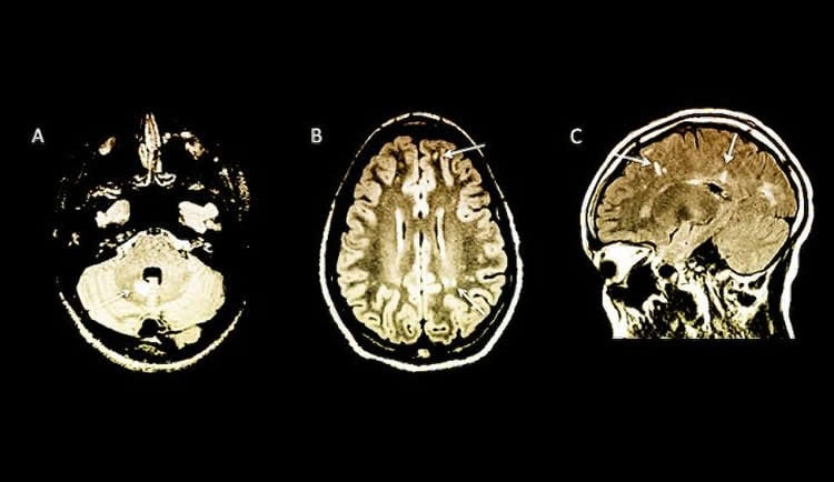
Published in the November issue of the journal Neurology: Neuroimmunology & Neuroinflammation, the study of 38 children at 16 sites in six countries showed that the MRIs can reveal changes in the brain associated with MS before the clinical symptoms of the disease appear in children.
The children in the study all underwent MRI scans for other reasons, most commonly headache, but the MRIs unexpectedly revealed signs of MS. Having MRI findings of MS without any symptoms of the disease has been termed radiologically isolated syndrome (RIS) and previously had only been seen in adults.
“For the first time we have proposed a definition of RIS in children,” said lead author Naila Makhani, M.D., assistant professor of pediatrics and neurology at Yale School of Medicine. “Children with RIS may represent a high-risk group of children that needs to be followed more closely for the later development of clinical multiple sclerosis.”
Approximately 42% of children in the study with MRI findings of MS developed the first clinical symptoms of the disease about two years after the abnormal MRI, which shows a faster development of the disease than has been reported in adults. Children who had a specific marker in spinal fluid or who had MRI changes in the spinal cord, were at greatest risk of developing the clinical symptoms of MS.
Makhani said five of the children in the study received an approved treatment for multiple sclerosis to try to prevent the disease. This number is too small to accurately draw conclusions about the effect of treatment, she noted.
“We hope that our work will help inform expert guidelines for how to follow up children with RIS and help us accurately inform families of the risk of later developing multiple sclerosis, something we were previously unable to do,” said Makhani.
Other authors on the study include Christine Lebrun, M.D.; Aksel Siva, M.D.; David Brassat M.D.; Clarisse Carra Dallière, M.D.; Jérôme de Seze, M.D.; Wei Du; Francoise Durand Dubief M.D.; Orhun Kantarci M.D.; Megan Langille, M.D.; Sona Narula M.D.; Jean Pelletier, M.D.; Juan Ignacio Rojas M.D.; Eugene D. Shapiro, M.D.; Robert T. Stone, M.D.; Mar Tintoré, M.D.; Ugur Uygunoglu M.D.; Patrick Vermersch, M.D.; Evangeline Wassmer M.D.; Darin T. Okuda, M.D.; and Daniel Pelletier, M.D.
Funding: The study was funded in part, by Yale’s Clinical and Translational Science Award from NIH (CTSA grant UL1TR000142 from the National Center for Advancing Translational Science [NCATS]).
Source: Karen N. Peart – Yale
Image Source: NeuroscienceNews.com image is credited to Yale.
Original Research: Full open access research for “Radiologically isolated syndrome in children: Clinical and radiologic outcomes” by Naila Makhani, MD, MPH, Christine Lebrun, MD, PhD, Aksel Siva, MD, David Brassat, MD, Clarisse Carra Dallière, MD, Jérôme de Seze, MD, Wei Du, MS, Françoise Durand Dubief, MD, Orhun Kantarci, MD, Megan Langille, MD, Sona Narula, MD, Jean Pelletier, MD, Juan Ignacio Rojas, MD, Eugene D. Shapiro, MD, Robert T. Stone, MD, Mar Tintoré, MD, Ugur Uygunoglu, MD, Patrick Vermersch, MD, Evangeline Wassmer, MD, Darin T. Okuda, MD and Daniel Pelletier, MD On behalf of Observatoire Francophone de la Sclérose en Plaques (OFSEP), Société Francophone de la Sclérose en Plaques (SFSEP), and the Radiologically Isolated Syndrome Consortium (RISC) in Neurology: Neuroimmunology & Neuroinflammation. Published online September 25 2017 doi:10.1212/NXI.0000000000000395
[cbtabs][cbtab title=”MLA”]Yale “Discovery in Amygdala Sheds Light on Emotional and Social Behavior Regulation.” NeuroscienceNews. NeuroscienceNews, 13 October 2017.
<https://neurosciencenews.com/multiple-sclerosis-mri-children-7740/>.[/cbtab][cbtab title=”APA”]Yale (2017, October 13). Discovery in Amygdala Sheds Light on Emotional and Social Behavior Regulation. NeuroscienceNews. Retrieved October 13, 2017 from https://neurosciencenews.com/multiple-sclerosis-mri-children-7740/[/cbtab][cbtab title=”Chicago”]Yale “Discovery in Amygdala Sheds Light on Emotional and Social Behavior Regulation.” https://neurosciencenews.com/multiple-sclerosis-mri-children-7740/ (accessed October 13, 2017).[/cbtab][/cbtabs]
Abstract
Radiologically isolated syndrome in children: Clinical and radiologic outcomes
Objective: To describe clinical and radiologic outcomes of children with incidental findings on neuroimaging suggestive of CNS demyelination (termed “radiologically isolated syndrome” or RIS).
Methods: Clinical and radiologic data were obtained from a historical cohort of children with no symptoms of demyelinating disease who had MRI scans that met the 2010 MRI criteria for dissemination in space for MS.
Results: We identified 38 children (27 girls and 11 boys) with RIS now being prospectively followed at 16 sites in 6 countries. The mean follow-up time was 4.8 ± 5.3 years. The most common reason for initial neuroimaging was headache (20/38, 53%). A first clinical event consistent with CNS demyelination occurred in 16/38 children (42%; 95% confidence interval [CI]: 27%–60%) in a median of 2.0 years (interquartile range [IQR] 1.0–4.3 years). Radiologic evolution developed in 23/38 children (61%; 95% CI: 44%–76%) in a median of 1.1 years (IQR 0.5–1.9 years). The presence of ≥2 unique oligoclonal bands in CSF (hazard ratio [HR] 10.9, 95% CI: 1.4–86.2, p = 0.02) and spinal cord lesions on MRI (HR 7.8, 95% CI: 1.4–43.6, p = 0.02) were associated with an increased risk of a first clinical event after adjustment for age and sex.
Conclusions: We describe the clinical characteristics and outcomes of children with incidental MRI findings highly suggestive of CNS demyelination. Children with RIS had a substantial risk of subsequent clinical symptoms and/or radiologic evolution. The presence of oligoclonal bands in CSF and spinal cord lesions on MRI were associated with an increased risk of a first clinical event.
“Radiologically isolated syndrome in children: Clinical and radiologic outcomes” by Naila Makhani, MD, MPH, Christine Lebrun, MD, PhD, Aksel Siva, MD, David Brassat, MD, Clarisse Carra Dallière, MD, Jérôme de Seze, MD, Wei Du, MS, Françoise Durand Dubief, MD, Orhun Kantarci, MD, Megan Langille, MD, Sona Narula, MD, Jean Pelletier, MD, Juan Ignacio Rojas, MD, Eugene D. Shapiro, MD, Robert T. Stone, MD, Mar Tintoré, MD, Ugur Uygunoglu, MD, Patrick Vermersch, MD, Evangeline Wassmer, MD, Darin T. Okuda, MD and Daniel Pelletier, MD On behalf of Observatoire Francophone de la Sclérose en Plaques (OFSEP), Société Francophone de la Sclérose en Plaques (SFSEP), and the Radiologically Isolated Syndrome Consortium (RISC) in Neurology: Neuroimmunology & Neuroinflammation. Published online September 25 2017 doi:10.1212/NXI.0000000000000395


