Summary: Researchers were able to predict the orientation preference of individual neurons by adding up the responses of their dendritic spines, a new study reports.
Source: Max Planck Florida.
New insights into neural computations in cerebral cortex.
The cerebral cortex is the largest and most complex area of the brain, comprising 20 billion neurons and 60 trillion synapses-a neuronal network whose proper function is critical for sensory perception, motor control, and cognition. The part of the cerebral cortex devoted to vision has played a key role in elucidating fundamental principles that are used by cortical circuits to encode information. Because edges supply a wealth of information about our visual world, neurons in visual cortex respond selectively to edge orientation, some preferring vertical edges, while others prefer horizontal, and all angles in between. Individual neurons also exhibit considerable diversity in their degree of selectivity, some responding to a narrow range of orientations, others to a broad range of orientations. These differences in selectivity are critical for accurately encoding the visual information in natural scenes, but the underlying mechanisms that account for this diversity remain unclear. In their recent publication in Nature Neuroscience, MPFI researchers Daniel Wilson, David Whitney, Ben Scholl and David Fitzpatrick describe how this diversity comes about and, in the process, provide new insights into the powerful role that dendrites play in cortical processing.
The research team addressed this issue using new microscopic imaging technologies that allowed them for the first time to assess the input/output functions of individual cortical neurons in the living brain. By using in vivo 2-photon calcium imaging, they were able to characterize the orientation tuning and spatial arrangement of synaptic inputs to the dendritic spines of individual neurons in ferret visual cortex, and compare dendritic spine and cell body responses.
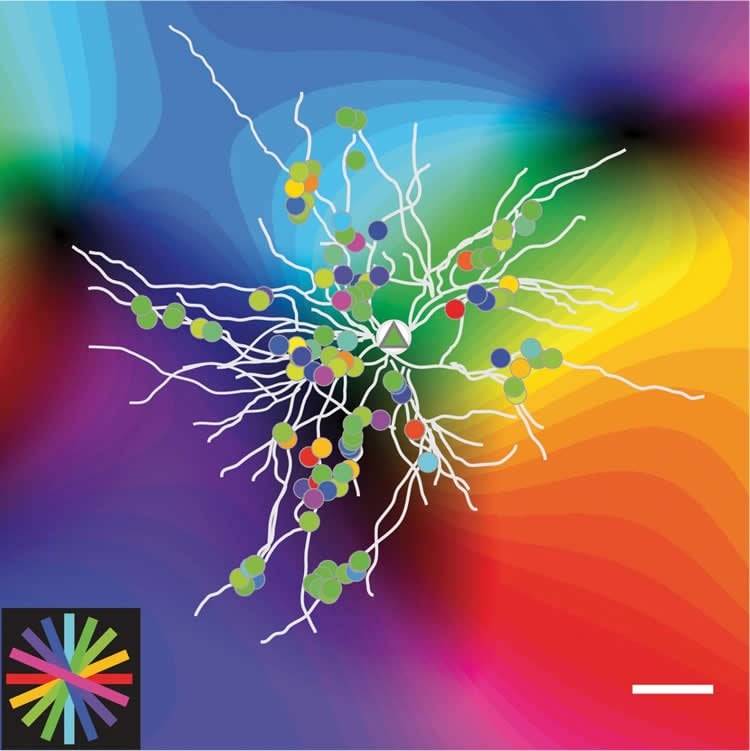
The researchers found that they were able to reliably predict the orientation preference of individual neurons simply by adding up the responses of their dendritic spines. However, the responses of the dendritic spines did not account for the degree of orientation selectivity exhibited by individual neurons. In looking for factors that could account for differences in selectivity, they noticed that spines with similar orientation preference were often spatially clustered along the dendrite and that neurons that had a greater number of these clusters exhibited greater selectivity. They also discovered that this functional clustering was correlated with localized dendritic events that are likely to enhance the inputs from the clustered spines.
There is more to dendrites than meets the eye: cluster synaptic inputs and orientation selectivity.
So not only did the researchers solve the riddle of orientation selectivity, they provided evidence that dendrites endow neurons with more computational power than previously thought. While this study focused specifically on information coding in visual cortex, it is likely that functional clustering of inputs within the dendritic field is a common principle influencing neuronal input/output functions throughout the cerebral cortex, significantly enhancing the brain’s information processing capabilities.
Facts:
- Dendrites are the tree-like extensions from neurons that serve as the main location for synapses–the junctions that neurons use to communicate with each other. In the cerebral cortex, dendrites are covered with thousands of small protrusions called spines and each dendritic spine is the site of a different synaptic input.
- The classical view of dendritic function is that information arriving via the dendritic spines is passively conveyed to the cell body through the dendrites, much like a cable, without changing the information content.
- Although it has been suggested that dendrites could play a more active role in this process, support for such a role in the functioning brain has been difficult to obtain.
- Now scientists at the Max Planck Florida Institute for Neuroscience (MPFI) have provided strong evidence that the arrangement of synaptic connections within the dendritic field supports an active role for dendrites in cortical processing and that these dendritic computations shape how neurons encode visual information.
Source: Jennifer Gutierrez – Max Planck Florida
Image Source: This NeuroscienceNews.com image is credited to Max Planck Florida Institute for Neuroscience.
Video Source: The video is credited to Max Planck Florida Institute for Neuroscience.
Original Research: Abstract for “Orientation selectivity and the functional clustering of synaptic inputs in primary visual cortex” by Daniel E Wilson, David E Whitney, Benjamin Scholl and David Fitzpatrick in Nature Neuroscience. Published online June 13 2016 doi:10.1038/nn.4323
[cbtabs][cbtab title=”MLA”]Max Planck Florida. “An Active Role for Dendrites in Cortical Processing.” NeuroscienceNews. NeuroscienceNews, 13 June 2016.
<https://neurosciencenews.com/dendrites-cortical-processing-4446/>.[/cbtab][cbtab title=”Max Planck Florida”]Max Planck Florida. (2016, June 13). An Active Role for Dendrites in Cortical Processing. NeuroscienceNews. Retrieved June 13, 2016 from https://neurosciencenews.com/dendrites-cortical-processing-4446/[/cbtab][cbtab title=”Chicago”]Max Planck Florida. “An Active Role for Dendrites in Cortical Processing.” https://neurosciencenews.com/dendrites-cortical-processing-4446/ (accessed June 13, 2016).[/cbtab][/cbtabs]
Abstract
Orientation selectivity and the functional clustering of synaptic inputs in primary visual cortex
The majority of neurons in primary visual cortex are tuned for stimulus orientation, but the factors that account for the range of orientation selectivities exhibited by cortical neurons remain unclear. To address this issue, we used in vivo two-photon calcium imaging to characterize the orientation tuning and spatial arrangement of synaptic inputs to the dendritic spines of individual pyramidal neurons in layer 2/3 of ferret visual cortex. The summed synaptic input to individual neurons reliably predicted the neuron’s orientation preference, but did not account for differences in orientation selectivity among neurons. These differences reflected a robust input–output nonlinearity that could not be explained by spike threshold alone and was strongly correlated with the spatial clustering of co-tuned synaptic inputs within the dendritic field. Dendritic branches with more co-tuned synaptic clusters exhibited greater rates of local dendritic calcium events, supporting a prominent role for functional clustering of synaptic inputs in dendritic nonlinearities that shape orientation selectivity.
“Orientation selectivity and the functional clustering of synaptic inputs in primary visual cortex” by Daniel E Wilson, David E Whitney, Benjamin Scholl and David Fitzpatrick in Nature Neuroscience. Published online June 13 2016 doi:10.1038/nn.4323