

Summary: The hippocampus and amygdala directly exchange signals in order to recognize emotional stimuli and encode them into memories, researchers report.
Source: UC Irvine.
Researchers at the University of California, Irvine have identified a key neural pathway in humans that explains how the brain processes feelings of fear and anxiety, a finding that could help scientists unlock new ways to treat mental health disorders.
People are motivated to remember fearful events, because this information is useful for daily survival. Yet over-interpretation of fear may lead to anxiety and other mental disorders. Understanding how the human brain processes fearful information has been a topic of intense scientific research. Until now, the brain circuit underlying fear has only been mapped in rodents.
The study, “Amygdala-hippocampal dynamics during salient information processing,” appears today in the journal Nature Communications.
Researchers recorded neuronal activity using electrodes inserted into the amygdala and hippocampus of nine people as they watch scenes from horror movies to stimulate the recognition of fear.
“Deep brain electrodes capture neurons firing millisecond by millisecond, revealing in real time how the brain attends to fearful stimuli,” said Jie Zheng, a UCI graduate student and the study’s first author.
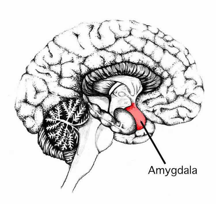
Researchers demonstrated that these two regions, nestled deep in the center of the brain and which play a key role in recognizing emotional stimuli and encoding them in memories, are directly exchanging signals.
“In fact, neurons in the amygdala fired 120 milliseconds earlier than the hippocampus. It is truly remarkable that we can measure the brain dynamics with such precision,” said Zheng. “Further, the traffic pattern between the two brain regions are controlled by the emotion of the movie; a unidirectional flow of information from the amygdala to the hippocampus only occurred when people were watching fearful movie clips but not while watching peaceful scenes.”
Human and animal studies have established the amygdala’s role in fear processing and a parallel role the hippocampus plays in enhanced memory processing of emotional events. Despite the breadth of this research, senior author Dr. Jack Lin, said it was not previously known how these two nearby brain regions interact during the recognition of fearful stimulus.
“Most studies focus on each brain region in isolation,” said Lin, a UCI professor of neurology. “Our study unifies the varied literature on the roles of the amygdala and hippocampus in emotional processing, with direct evidence that the amygdala first extracts emotional relevance and then sends this information to the hippocampus to be processed as a memory.”
Understanding the activation of the exact brain network in processing fearful stimuli is critical to develop new treatment for psychiatric disorders in the era of personalized medicine.
“This is the first study in humans to delineate the mechanism by which our brain processes fear at the circuitry level,” Lin said. “This has huge implications for treating neuropsychiatric disorders. For example, current drugs available to treat anxiety disorder bind to large areas of the brain, leading to unwanted side effects.” “Our hope is that we will one day be able to target and manipulate the precise amygdala-hippocampal circuit involved in processing negative emotions while preserving positive ones,” he said. “This study brings the promise of targeted therapy a step closer.”
Measurements were collected from electrodes implanted by UC Irvine Health neurosurgeons in nine patients with medication-resistant epilepsy as part of an assessment of their seizure activity. Electrode placement was guided exclusively by these patients’ clinical needs, Lin said
The study was conducted in collaboration with Robert Knight, a UC Berkeley professor of psychology; postdoctoral fellow Kristopher Anderson; and research scientist Avgusta Shestyuk. The deep brain electrodes were placed by UC Irvine Health neurosurgeons Dr. Frank Hsu and Dr. Sumeet Vadera. UC Irvine Health neurologist Dr. Lilit Mnatsakanyan recruited patients for the study. Gultekin Gulsen, UCI associate professor of radiology and biomedical engineering, provided analysis tools. Michael Yassa, UCI associate professor of neurobiology and behavior, and postdoctoral fellow Stephanie Leal helped design the study.
Funding: This study was supported with funding from the National Institute of Neurological Disorders and Stroke awards 2R37 NS021135 and K23 NS060993, National Institute on Deafness and Other Communication Disorders award R01 007293, the Nielsen Corporation and the UCI School of Medicine Bridge Fund.
Source: John Murray – UC Irvine
Image Source: NeuroscienceNews.com image is in the public domain.
Original Research: Full open access research for “Amygdala-hippocampal dynamics during salient information processing” by Jie Zheng, Kristopher L. Anderson, Stephanie L. Leal, Avgusta Shestyuk, Gultekin Gulsen, Lilit Mnatsakanyan, Sumeet Vadera, Frank P. K. Hsu, Michael A. Yassa, Robert T. Knight & Jack J. Lin in Nature Communications. Published online February 8 2017 doi:10.1038/ncomms14413
[cbtabs][cbtab title=”MLA”]UC Irvine “Horror Movie Scenes Help Researchers Identify Key Brain Circuits for Fear Processing.” NeuroscienceNews. NeuroscienceNews, 8 February 2017.
<https://neurosciencenews.com/amygdala-hippocampus-fear-movies-6082/>.[/cbtab][cbtab title=”APA”]UC Irvine (2017, February 8). Horror Movie Scenes Help Researchers Identify Key Brain Circuits for Fear Processing. NeuroscienceNew. Retrieved February 8, 2017 from https://neurosciencenews.com/amygdala-hippocampus-fear-movies-6082/[/cbtab][cbtab title=”Chicago”]UC Irvine “Horror Movie Scenes Help Researchers Identify Key Brain Circuits for Fear Processing.” https://neurosciencenews.com/amygdala-hippocampus-fear-movies-6082/ (accessed February 8, 2017).[/cbtab][/cbtabs]
Abstract
Amygdala-hippocampal dynamics during salient information processing
Recognizing motivationally salient information is critical to guiding behaviour. The amygdala and hippocampus are thought to support this operation, but the circuit-level mechanism of this interaction is unclear. We used direct recordings in the amygdala and hippocampus from human epilepsy patients to examine oscillatory activity during processing of fearful faces compared with neutral landscapes. We report high gamma (70–180 Hz) activation for fearful faces with earlier stimulus evoked onset in the amygdala compared with the hippocampus. Attending to fearful faces compared with neutral landscape stimuli enhances low-frequency coupling between the amygdala and the hippocampus. The interaction between the amygdala and hippocampus is largely unidirectional, with theta/alpha oscillations in the amygdala modulating hippocampal gamma activity. Granger prediction, phase slope index and phase lag analysis corroborate this directional coupling. These results demonstrate that processing emotionally salient events in humans engages an amygdala-hippocampal network, with the amygdala influencing hippocampal dynamics during fear processing.
“Amygdala-hippocampal dynamics during salient information processing” by Jie Zheng, Kristopher L. Anderson, Stephanie L. Leal, Avgusta Shestyuk, Gultekin Gulsen, Lilit Mnatsakanyan, Sumeet Vadera, Frank P. K. Hsu, Michael A. Yassa, Robert T. Knight & Jack J. Lin in Nature Communications. Published online February 8 2017 doi:10.1038/ncomms14413


