Summary: Researchers shed light on how the eyes compute the direction of moving light.
Source: University of Queensland.
The mystery of how human eyes compute the direction of moving light has been made clearer by scientists at The University of Queensland.
Using advanced electrical recording techniques, researchers from UQ’s Queensland Brain Institute (QBI) discovered how nerve cells in the eye’s retina were integral to the process.
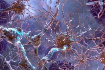
Professor Stephen Williams said that dendrites – the branching processes of a neuron that conduct electrical signals toward the cell body – played a critical role in decoding images.
“The retina is not a simple camera, but actively processes visual information in a neuronal network, to compute abstractions that are relayed to the higher brain,” Professor Williams said.
“Previously, dendrites of neurons were thought to be passive input areas.
“Our research has found that dendrites also have powerful processing capabilities.”
Co-author Dr Simon Kalita-de Croft said dendritic processing enabled the retina to convert and refine visual cues into electrical signals.
“We now know that movement of light – say, a flying bird, or a passing car – gets converted into an electrical signal by dendritic processing in the retina,” Dr Kalita-de Croft said.

“The discovery bridges the gap between our understanding of the anatomy and physiology of neuronal circuits in the retina.”
Professor Williams said the ability of dendrites in the retina to process visual information depended on the release of two neurotransmitters – chemical messengers – from a single class of cell.
“These signals are integrated by the output neurons of the retina,” Professor Williams said.
“Determining how the neural circuits in the retina process information can help us understand computational principles operational throughout the brain.
“Excitingly, our discovery provides a new template for how neuronal computations may be implemented in brain circuits.”
Source: Donna Lu – University of Queensland
Image Source: NeuroscienceNews.com image is in the public domain.
Original Research: Full open access research for “Dendro-dendritic cholinergic excitation controls dendritic spike initiation in retinal ganglion cells” by A. Brombas, S. Kalita-de Croft, E. J. Cooper-Williams & S. R. Williams in Nature Communications. Published online June 7 2017 doi:10.1038/ncomms15683
[cbtabs][cbtab title=”MLA”]University of Queensland “Shedding Light on How Our Brain Processes Visual Cues.” NeuroscienceNews. NeuroscienceNews, 7 June 2017.
<https://neurosciencenews.com/visual-cue-eye-processing-6856/>.[/cbtab][cbtab title=”APA”]University of Queensland (2017, June 7). Shedding Light on How Our Brain Processes Visual Cues. NeuroscienceNew. Retrieved June 7, 2017 from https://neurosciencenews.com/visual-cue-eye-processing-6856/[/cbtab][cbtab title=”Chicago”]University of Queensland “Shedding Light on How Our Brain Processes Visual Cues.” https://neurosciencenews.com/visual-cue-eye-processing-6856/ (accessed June 7, 2017).[/cbtab][/cbtabs]
Abstract
Dendro-dendritic cholinergic excitation controls dendritic spike initiation in retinal ganglion cells
The retina processes visual images to compute features such as the direction of image motion. Starburst amacrine cells (SACs), axonless feed-forward interneurons, are essential components of the retinal direction-selective circuitry. Recent work has highlighted that SAC-mediated dendro-dendritic inhibition controls the action potential output of direction-selective ganglion cells (DSGCs) by vetoing dendritic spike initiation. However, SACs co-release GABA and the excitatory neurotransmitter acetylcholine at dendritic sites. Here we use direct dendritic recordings to show that preferred direction light stimuli evoke SAC-mediated acetylcholine release, which powerfully controls the stimulus sensitivity, receptive field size and action potential output of ON-DSGCs by acting as an excitatory drive for the initiation of dendritic spikes. Consistent with this, paired recordings reveal that the activation of single ON-SACs drove dendritic spike generation, because of predominate cholinergic excitation received on the preferred side of ON-DSGCs. Thus, dendro-dendritic release of neurotransmitters from SACs bi-directionally gate dendritic spike initiation to control the directionally selective action potential output of retinal ganglion cells.
“Dendro-dendritic cholinergic excitation controls dendritic spike initiation in retinal ganglion cells” by A. Brombas, S. Kalita-de Croft, E. J. Cooper-Williams & S. R. Williams in Nature Communications. Published online June 7 2017 doi:10.1038/ncomms15683


