

The discovery has implications for understanding how the human brain evolved and how it varies between people.
When you stare in puzzlement at an optical illusion, two distinct parts of the neocortex in your brain are hard at work: the primary visual cortex is receiving information on what your eyes see, and the surrounding higher order visual areas are trying to interpret that tricky amalgam of information. These two areas, though, are linked in more ways than just function—the same gene controls the size of each area, Salk researchers led by Dennis O’Leary have now discovered.
The findings, published in the journal eLife, help shed light on how the human brain has evolved and could explain why some people are better at seeing optical illusions than others.
The neocortex—that outermost, wrinkly layer—of the mammalian brain has primary and higher order areas for vision, hearing and touch. In each case, the primary areas receive sensory information from the thalamus and the higher order areas process that information. Scientists have long assumed that the sizes of these areas were independent of each other—in part because primates have larger higher order areas compared to other animals.
About a decade ago, O’Leary’s research group discovered that a gene called Emx2 controlled the size of the primary visual area of the neocortex. Since, the lab has studied the development and organization of the visual cortex by inactivating Emx2. Most recently, the team wondered what happened to higher order areas when the primary areas were changed.
“We wanted to know how higher order areas are affected when the visual cortex is unusually large or unusually small to better understand how the layout of the cortex is generated during development,” says Andreas Zembrzycki, a senior research associate in the O’Leary lab and first author of the paper.
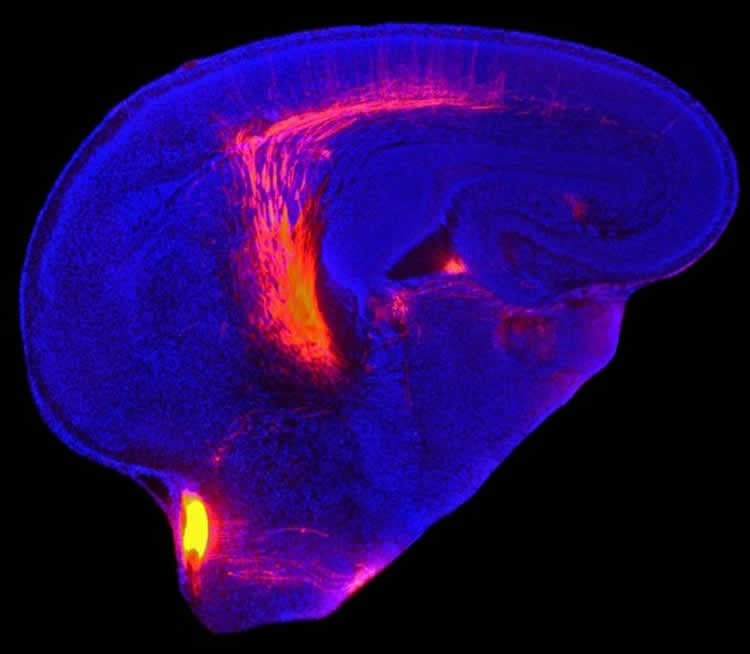
Zembrzycki and O’Leary turned up or down the activity of Emx2 in mice to make the primary visual areas larger or smaller. They then measured the size of the higher order visual areas. In each case, the higher order areas grew or shrank in direct proportion to the primary areas—when the primary area was 150 percent larger, for instance, the higher order areas were also about one and a half times larger.
“This was really a very big surprise,” says Zembrzycki. The finding indicates that, throughout evolution, higher order structures likely didn’t grow larger out of proportion with corresponding primary areas. The new results also suggest that people with larger primary sensory orders likely have larger corresponding higher order areas. That’s important because there’s a huge variability—up to 300 percent—in the sizes of the primary visual areas of humans.
Recently, scientists discovered that people with smaller primary visual areas were fooled more often and more strongly by optical illusions. Likewise, the larger your primary visual area, the better you are at interpreting optical illusions.
“Since the size of the primary area and size of the higher order areas are linked we expect that these people also have larger high order visual areas and that’s what helping them see the illusion clearly,” says Zembrzycki. Aside from the general population, perturbations in the size and structure of the primary sensory areas have been linked to diseases including autism. “In light of our findings, it’s definitely worth considering that higher order area deficits might also contribute to disease,” he adds.
Since the current work was done in mice, O’Leary and Zembrzycki want to confirm the link in humans by using brain scans to measure the natural variation in the neocortical areas and search for potential links to disease.
Other researchers on the study were Adam M. Stocker, Axel Leingartner, Setsuko Sahara, Shen-Ju Chou and Scott May of the Salk Institute; and Valery Kalatsky and Michael Stryker of the University of California, San Francisco.
Funding: The work was supported by the Vincent J. Coates Chair of Molecular Neurobiology at the Salk Institute for Biological Sciences.
Source: Salk Institute
Image Source: The image is credited to Salk Institute.
Original Research: Full open access research “Genetic mechanisms control the linear scaling between related cortical primary and higher order sensory areas” by Andreas Zembrzycki, Adam M Stocker, Axel Leingärtner, Setsuko Sahara, Shen-Ju Chou, Valery Kalatsky, Scott R May, Michael P Stryker, and Dennis DM O’Leary in eLife. Published online December 24 2015 doi:10.7554/eLife.11416
Abstract
Genetic mechanisms control the linear scaling between related cortical primary and higher order sensory areas
In mammals, the neocortical layout consists of few modality-specific primary sensory areas and a multitude of higher order ones. Abnormal layout of cortical areas may disrupt sensory function and behavior. Developmental genetic mechanisms specify primary areas, but mechanisms influencing higher order area properties are unknown. By exploiting gain-of and loss-of function mouse models of the transcription factor Emx2, we have generated bi-directional changes in primary visual cortex size in vivo and have used it as a model to show a novel and prominent function for genetic mechanisms regulating primary visual area size and also proportionally dictating the sizes of surrounding higher order visual areas. This finding redefines the role for intrinsic genetic mechanisms to concomitantly specify and scale primary and related higher order sensory areas in a linear fashion.
“Genetic mechanisms control the linear scaling between related cortical primary and higher order sensory areas” by Andreas Zembrzycki, Adam M Stocker, Axel Leingärtner, Setsuko Sahara, Shen-Ju Chou, Valery Kalatsky, Scott R May, Michael P Stryker, and Dennis DM O’Leary in eLife. Published online December 24 2015 doi:10.7554/eLife.11416


