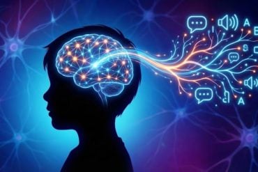
Summary: According to a new study, the brain activity of those who dream during NREM sleep is closer to the activity of awake people, compared to those who do not dream.
Source: Aalto University.
Measurements demonstrated that the brain activity of people who dream during NREM sleep, compared to people who do not dream, is closer to the brain activity of awake people.
Researchers from Aalto University and the University of Wisconsin utilised a TMS-EEG device, which combines transcranial magnetic stimulation and EEG, to examine how the brain activity of people in the restful non-rapid eye movement (NREM) sleep is affected by whether they dream or do not dream.
When the NREM sleep of subjects had lasted at least three minutes, researchers gave magnetic pulses that induced a weak electric field and activated neurons. After a series of pulses, the subject was woken with an alarm sound, and they were then asked whether they had dreamed and to describe the content of the dream.
‘It is traditionally thought that dreaming occurs only in REM sleep. However, as also our study demonstrates, subjects woken from NREM sleep are also able to give accounts of their dreams in more than half of cases,’ Post-doctoral Researcher Jaakko Nieminen from Aalto University explains.
‘EEG showed that the deterministic brain activity produced by magnetic pulses was notably shorter in people who did not dream, i.e. were unconscious, than in people who had dreamt. We also observed that the longer the story about the dream, the more the subject’s EEG resembled that measured from people who were awake,’ Dr Nieminen explains.
Assessment of consciousness may help in treatment of brain injury patients
Dr Nieminen performed the measurements with his research colleague Olivia Gosseries at the University of Wisconsin-Madison Center for Sleep and Consciousness, which is headed by Giulio Tononi one of the world’s most renowned researchers of consciousness. The measurements were carried out during a period of over 40 nights and a total of 11 subjects participated. Due to sleeping difficulties and other challenges, reliable measurements could only be acquired from six subjects. During the night, subjects were woken a maximum of 16 times.

‘Consciousness in different physiological states (e.g. during wakefulness, sleep, anesthesia and vegetative state) has previously been researched with TMS-EEG measurements. We wanted to eliminate all other differences related to the different states as thoroughly as possible, and for this reason we focused on the narrow physiological state of NREM sleep,’ Dr Nieminen notes.
Transcranial magnetic stimulation is already utilised in such things as the treatment of depression and pain. According to Dr Nieminen, in the future the precise data provided by TMS¬-EEG measurements on the state of consciousness may also help e.g. in the treatment of those brain injury patients who are unable to communicate.
Source: Jaakko Nieminen – Aalto University
Image Source: This NeuroscienceNews.com image is credited to Aalto University and University of Wisconsin.
Original Research: Full open access research for “Consciousness and cortical responsiveness: a within-state study during non-rapid eye movement sleep” by Jaakko O. Nieminen, Olivia Gosseries, Marcello Massimini, Elyana Saad, Andrew D. Sheldon, Melanie Boly, Francesca Siclari, Bradley R. Postle and Giulio Tononi in Scientific Reports. Published online August 5 2016 doi:10.1038/srep30932
[cbtabs][cbtab title=”MLA”]Aalto University. “Measuring Differences in Brain Activity of Those Who Dream and Those Who Don’t.” NeuroscienceNews. NeuroscienceNews, 9 August 2016.
<https://neurosciencenews.com/tms-eeg-dream-neural-activity-4813/>.[/cbtab][cbtab title=”APA”]Aalto University. (2016, August 9). Measuring Differences in Brain Activity of Those Who Dream and Those Who Don’t. NeuroscienceNews. Retrieved August 9, 2016 from https://neurosciencenews.com/tms-eeg-dream-neural-activity-4813/[/cbtab][cbtab title=”Chicago”]Aalto University. “Measuring Differences in Brain Activity of Those Who Dream and Those Who Don’t.” https://neurosciencenews.com/tms-eeg-dream-neural-activity-4813/ (accessed August 9, 2016).[/cbtab][/cbtabs]
Abstract
Consciousness and cortical responsiveness: a within-state study during non-rapid eye movement sleep
When subjects become unconscious, there is a characteristic change in the way the cerebral cortex responds to perturbations, as can be assessed using transcranial magnetic stimulation and electroencephalography (TMS–EEG). For instance, compared to wakefulness, during non-rapid eye movement (NREM) sleep TMS elicits a larger positive–negative wave, fewer phase-locked oscillations, and an overall simpler response. However, many physiological variables also change when subjects go from wake to sleep, anesthesia, or coma. To avoid these confounding factors, we focused on NREM sleep only and measured TMS-evoked EEG responses before awakening the subjects and asking them if they had been conscious (dreaming) or not. As shown here, when subjects reported no conscious experience upon awakening, TMS evoked a larger negative deflection and a shorter phase-locked response compared to when they reported a dream. Moreover, the amplitude of the negative deflection—a hallmark of neuronal bistability according to intracranial studies—was inversely correlated with the length of the dream report (i.e., total word count). These findings suggest that variations in the level of consciousness within the same physiological state are associated with changes in the underlying bistability in cortical circuits.
“Consciousness and cortical responsiveness: a within-state study during non-rapid eye movement sleep” by Jaakko O. Nieminen, Olivia Gosseries, Marcello Massimini, Elyana Saad, Andrew D. Sheldon, Melanie Boly, Francesca Siclari, Bradley R. Postle and Giulio Tononi in Scientific Reports. Published online August 5 2016 doi:10.1038/srep30932


