Feeding is essential for survival. Senses such as smell or sight can help guide us to good food sources, but the final decision to eat or reject a potential food is controlled by taste.
While the tongue is the main taste organ in humans and other mammals, insects not only have taste organs inside their mouthparts, but also on the body. Their legs, wings and ovipositor (the organ with which females lay eggs) can contain taste receptor neurons.
The need for so many taste organs in insects is not understood very well. However, a clue for understanding their function comes from examining the anatomy of the taste-sensing neurons. These neurons send projections to different parts of the central nervous system of the fly. This suggests that taste information received from different parts of the body is processed differently in the brain. Therefore, different taste organs may have different functions.
To test this idea, Thoma et al. used the fruit fly Drosophila as a model for insects. With Drosophila, it is possible to target small numbers of neurons and to block them or activate them with genetic tools. The scientists blocked different groups of sweet-sensing neurons and measured sugar choice, the first step in feeding behavior. Normally, hungry flies choose sugar very quickly, but the flies which had all sweet-sensing neurons in their legs blocked could not choose sugar.
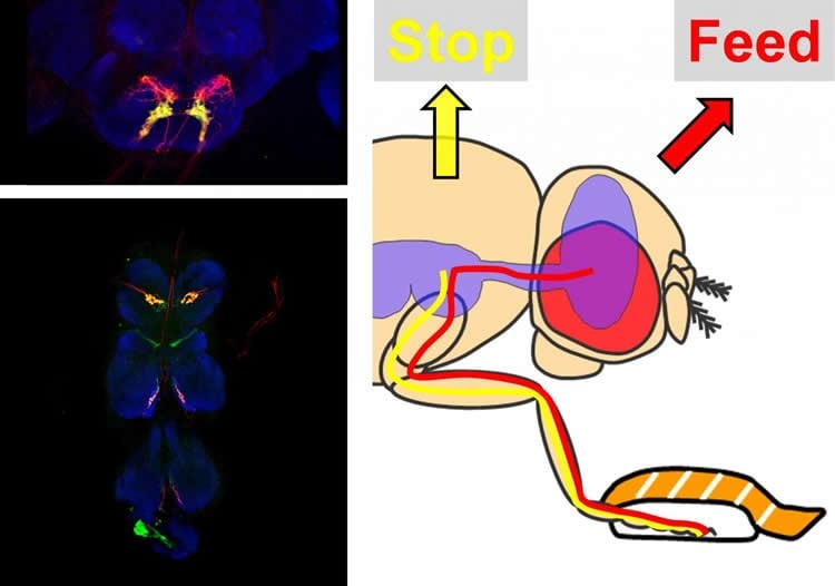
The scientists then examined the sweet taste neurons in the legs and found two populations of neurons. One group of neurons connected directly to the brain of the fly. The remaining neurons connected to the ventral nerve cord, a structure analogous to the spinal cord in humans.
The authors blocked each population separately and found that they have specialized functions. While the brain-projecting neurons are important for the initiation of feeding, the ventral nerve cord-projecting neurons are important for stopping the fly’s movement as soon as it steps on food. Both of these functions are important for correctly choosing sugar.
These results show how feeding, a complex behavior with many steps, is organized by the specialized contributions of different groups of neurons. The role of the taste neurons in other organs is only partially understood, but it is possible that they are important for the later stages of feeding.
Additionally, popular insect repellents such as DEET rely on chemicals that smell and taste bad to insects. Therefore, a better understanding of insect choice behavior and feeding may also contribute to the development of more effective pest control.
Source: Hiromu Tanimoto – Tohoku University
Image Credit: The image is credited to Vladimiros Thoma.
Original Research: Full open access research for “Functional dissociation in sweet taste receptor neurons between and within taste organs of Drosophila” by Vladimiros Thoma, Stephan Knapek, Shogo Arai, Marion Hartl, Hiroshi Kohsaka, Pudith Sirigrivatanawong, Ayako Abe, Koichi Hashimoto and Hiromu Tanimoto in Nature Communications. Published online February 19 2016 doi:10.1038/ncomms10678
Abstract
Functional dissociation in sweet taste receptor neurons between and within taste organs of Drosophila
Finding food sources is essential for survival. Insects detect nutrients with external taste receptor neurons. Drosophila possesses multiple taste organs that are distributed throughout its body. However, the role of different taste organs in feeding remains poorly understood. By blocking subsets of sweet taste receptor neurons, we show that receptor neurons in the legs are required for immediate sugar choice. Furthermore, we identify two anatomically distinct classes of sweet taste receptor neurons in the leg. The axonal projections of one class terminate in the thoracic ganglia, whereas the other projects directly to the brain. These two classes are functionally distinct: the brain-projecting neurons are involved in feeding initiation, whereas the thoracic ganglia-projecting neurons play a role in sugar-dependent suppression of locomotion. Distinct receptor neurons for the same taste quality may coordinate early appetitive responses, taking advantage of the legs as the first appendages to contact food.
“Functional dissociation in sweet taste receptor neurons between and within taste organs of Drosophila” by Vladimiros Thoma, Stephan Knapek, Shogo Arai, Marion Hartl, Hiroshi Kohsaka, Pudith Sirigrivatanawong, Ayako Abe, Koichi Hashimoto and Hiromu Tanimoto in Nature Communications. Published online February 19 2016 doi:10.1038/ncomms10678