Summary: It has long been a clinical observation that psychological stress leads to skin “flares,” but the biological “why” has remained elusive. A new study has finally mapped the direct neural highway between the mind and the skin.
By analyzing data from 51 patients and mouse models, researchers identified a specific set of sympathetic neurons that carry stress signals from the brain directly to the skin, where they recruit and “switch on” inflammatory immune cells.
Key Facts & Statistics
- The “Pdyn+” Pathway: Researchers identified a specific subset of prodynorphin-positive (Pdyn+) noradrenergic sympathetic neurons. These neurons act as the physical link between the brain’s stress response and the skin’s immune system.
- Hairy Skin Vulnerability: These specific neurons preferentially innervate hairy skin, making those areas more reactive to emotional distress.
- Eosinophil Recruitment: Stress signals use the CCL11–CCR3 signaling pathway to call eosinophils (inflammatory white blood cells) into the skin.
- The “On” Switch: Once in the skin, these immune cells are activated via beta-2 adrenergic receptors, releasing the proteins and cytokines that cause the itching and redness of atopic dermatitis.
- Therapeutic Potential: Genetic “ablation” (removal) of these neurons or the eosinophils themselves completely stopped stress-induced inflammation in models, suggesting that managing the nervous system is as vital as treating the skin surface.
Source: AAAS
New research leveraging patient data and mouse models reveals how psychological stress can worsen atopic dermatitis, or eczema. It does so by activating a specific neural pathway that links the brain to immune responses in the skin.
The study’s authors emphasize that managing psychological stress, alongside conventional therapies, may represent an underused but potentially powerful strategy for improving outcomes in eczema.

“[The authors] offer a mechanistic explanation for the well-documented but poorly understood link between stress and atopic dermatitis flare-ups,” write Nicolas Gaudenzio and Lillan Basso in a related Perspective.
“Research into the existence of similar mechanisms in other stress-sensitive inflammatory diseases, such as psoriasis or inflammatory bowel disease, is also needed,” they say.
Psychological stress is known to disrupt the body’s immune balance. The skin is particularly vulnerable because it contains dense networks of nerves and immune cells, making it highly responsive to stress-related signals. Conditions such as eczema illustrate this neurobiological connection, as stress frequently exacerbates the condition.
Recent studies suggest that stress signals transmitted through the sympathetic nervous system may directly influence immune activity in the skin. Moreover, eosinophils – immune cells that release inflammatory proteins and cytokines – are closely linked to dermatitis severity.
However, the mechanisms by which stress-driven neural signals recruit and activate these cells remain poorly understood.
To address this gap, Jiahe Tian and colleagues analyzed clinical data from 51 eczema patients and complementary mouse models to examine the relationship between stress and inflammatory immune responses in the skin.
Tian et al. identified a subset of prodynorphin-positive (Pdyn)+ noradrenergic sympathetic neurons that preferentially innervate hairy skin. Using genetic ablation and optogenetic activation, the authors tested how these neurons influence inflammation and eosinophil activity and found that higher stress in patients correlated with increased eosinophil accumulation in the skin.
In mouse models, Tian et al. show that Pdyn+ sympathetic neurons transmitted stress signals from the brain to the skin, worsening inflammation.
These neurons recruited eosinophils through the CCL11–CCR3 signaling pathway and activated them through the beta-2 adrenergic receptor. Removing either these neurons or eosinophils reduced stress-induced inflammation, while activating the neurons increased it.
Key Questions Answered:
A: Absolutely not. This study proves that your eczema is a very real neuro-immune event. Your brain isn’t “imagining” the itch; it is physically sending a command through a specific nerve wire (the Pdyn+ neuron) to your skin, telling your immune cells to attack. Stress is the “trigger,” but the inflammation in your skin is a physical biological response.
A: When you are stressed, your sympathetic nervous system (the “fight or flight” system) kicks into high gear. This research shows that those Pdyn+ neurons release chemicals that act like a “magnet” (CCL11) for eosinophils. These cells rush to your skin and release inflammatory proteins, creating that sudden, intense itch.
A: While relaxation and stress management are powerful, the study suggests we need a two-pronged approach. Traditional creams treat the skin, but we may eventually need “neuro-immune” therapies that block the specific beta-2 adrenergic receptors or the CCL11 pathway to stop the brain’s stress signal from reaching the skin in the first place.
Editorial Notes:
- This article was edited by a Neuroscience News editor.
- Journal paper reviewed in full.
- Additional context added by our staff.
About this stress and neuroscience research news
Author: Science Press Package Team
Source: AAAS
Contact: Science Press Package Team – AAAS
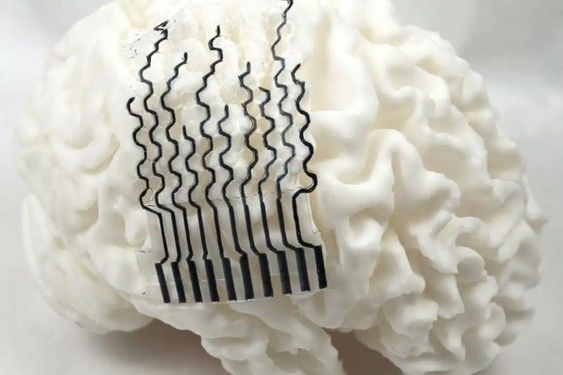
Image: The image is credited to Neuroscience News
Original Research: Closed access.
“A sympathetic-eosinophil axis orchestrates psychological stress to exacerbate skin inflammation” by Jiahe Tian, Yudian Cao, Yilei Li, Junlong Sun, Cheng Zhan, Wei Ni, Yongjun Zheng, Yanqing Wang, and Shenbin Liu. Science
DOI:10.1126/science.adv5974
Abstract
A sympathetic-eosinophil axis orchestrates psychological stress to exacerbate skin inflammation
INTRODUCTION
Psychological stress is a well-established exacerbating factor for atopic dermatitis (AD), capable of worsening inflammation through complex interactions among the nervous, endocrine, and immune systems. Stress hormones can directly compromise the skin barrier, promote inflammation, and intensify the sensation of itch.
Scratching in response to itch further damages the skin, creating a self-perpetuating cycle of inflammation and psychological distress. Consequently, stress management is considered an integral component in the comprehensive management of AD.
RATIONALE
Eosinophilic infiltration is a pathological hallmark of AD. Eosinophil-derived mediators, including granule proteins such as eosinophil peroxidase (Epx) and major basic protein (MBP), as well as cytokines such as interleukin-31 (IL-31), potently amplify inflammatory cascades and correlate with disease severity.
The mechanistic basis governing eosinophil recruitment and activation specifically within the context of psychological stress remains poorly understood, representing a significant knowledge gap in cutaneous neuroimmune communications.
Additionally, whereas sympathetic neurons exhibit target-specific organization and participate in peripheral immunomodulation during stress responses, the precise pathways through which psychological stress converges with atopy-associated inflammation remain incompletely delineated, particularly regarding their connection to eosinophil-mediated inflammation in stressed skin.
RESULTS
To determine the immune mediators and neural pathways through which stress signals aggravate skin inflammation, we conducted a retrospective analysis of AD patients and studied stress-challenged murine AD models. Our analysis revealed a specific association between stress-induced eosinophilia and skin inflammation severity in AD patients.
In mice, genetic ablation of eosinophils (in EpxiCre-DTA mice) conferred protection against stress-exacerbated dermatitis. Through chemical sympathectomy with 6-hydroxydopamine and surgical removal of the adrenal glands, we determined that peripheral sympathetic nerves, rather than the hypothalamus-pituitary-adrenal axis, mediates the stress-induced worsening of skin inflammation.
Using single-nucleus RNA sequencing and intersectional genetic approaches, we further identified two major populations of noradrenergic sympathetic neurons in mice, defined by prodynorphin (Pdyn) and neuropeptide Y (Npy) expression, that were activated by psychological stress.
Our functional studies suggested that skin-innervating Pdyn+ sympathetic neurons, but not their Npy+ counterparts, were both necessary and sufficient for driving stress-induced dermatitis and eosinophilia. Optogenetic activation of Pdyn+ neurons promoted eosinophil recruitment and exacerbated inflammation, effects that were abolished upon eosinophil depletion.
These Pdyn+ neurons were found to release the chemokine C-C motif ligand 11 (CCL11), which acts on its receptor, C-C chemokine receptor type 3 (CCR3), to mediate eosinophil chemotaxis. Finally, adrenergic signaling through adrenergic receptor beta2 (Adrb2) on eosinophils was critical, because eosinophil-specific Adrb2 knockout mitigated stress-induced exacerbation of dermatitis.
CONCLUSION
Our findings suggest that psychological stress exacerbates AD-like inflammation through a specialized subset of skin-innervating Pdyn+ noradrenergic sympathetic neurons that engage eosinophils through the CCL11-CCR3 chemotactic axis and Adrb2-mediated activation. These results indicate that stress-induced eosinophilia could be a potential biomarker of AD severity and suggest that targeting the Pdyn+ sympathetic neuron-eosinophil interface may offer therapeutic benefit in mitigating the inflammatory sequelae of psychological stress.