Summary: The postrhinal cortex of rats contains three types of spatial cells which act together to provide a sense of location and directional orientation.
Source: Dartmouth College
As you enter a new environment such as visiting a classroom for the first time, your brain takes in information about your surroundings to help inform where you are and what direction you are facing. Knowing where the center of the room is located helps provide a reference point for processing space. A Dartmouth study published in Science provides new insight into navigation and spatial learning by examining how the rat brain processes spatial information from the outside world in an egocentric framework and converts it to information in relation to the animal’s spatial position in an allocentric framework (referenced to the world at-large).
As the study explains, the postrhinal cortex of the rat brain is considered “the rodent homolog of the human parahippocampal cortex,” which is thought to be responsible for spatial navigation. The study examined how the rat brain processes incoming sensory information to inform where it is. For each trial, the rat was placed in an open box (“room”) and trained to forage for sugar pellets that fell from overhead. The rat was filmed and its brain activity was recorded using electrodes. Following the trials, the researchers analyzed what the spatial cells were doing in relation to where the rat was in the environment. The results demonstrated that the postrhinal cortex contains three types of spatial cells, which act together to provide a sense of where the rat is located and its directional orientation within the local environment.
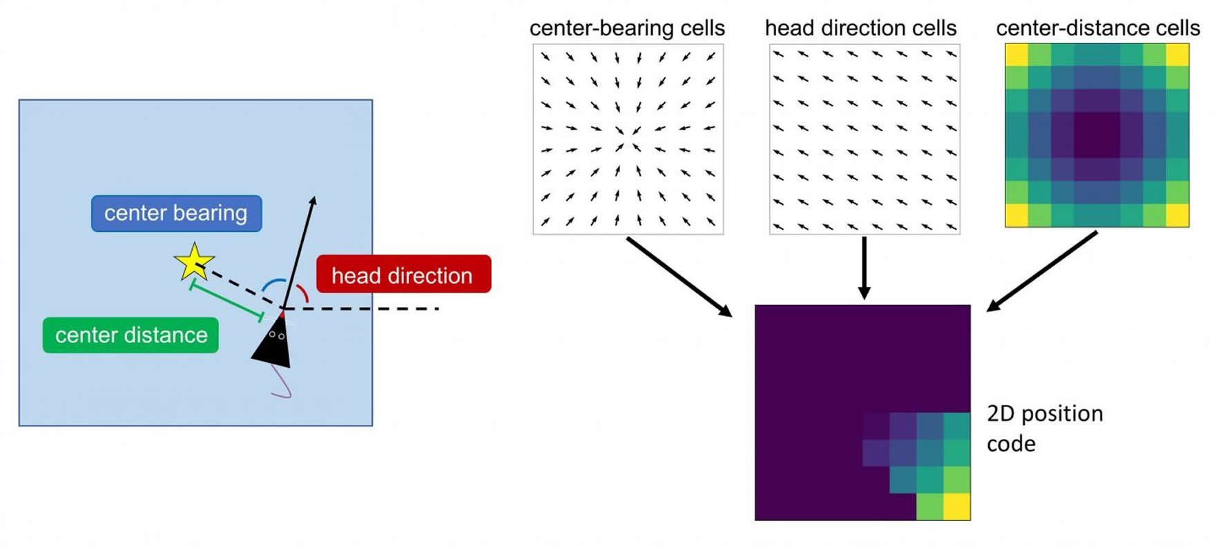
- Some neurons are “center-bearing” cells, which are egocentric in nature, and identify where the geometric center of the space is located in relation to the animal. The center-bearing cells inform the animal of its directional orientation relative to where the center of room is, such as whether the room center is in front or behind the animal.
- Other neurons are “center-distance” cells, whose firing rates encode the distance the rat is from the center of the environment. Some center-distance cells may fire more rapidly in the center while others may fire more slowly there, but both types of these cells are encoding the same type of information.
- Finally, other neurons are “head-direction” cells, which respond to and are specific to a cardinal direction. Regardless of where the animal is located in the room, the cell will fire whenever the animal is looking in a specific direction. Different head direction cells encode different directions and the population of this cell type appears to encode all 360 degrees.
Together, the center-bearing cells, center-distance cells and head-direction cells tell the animal where it is and what direction it is facing.
“Our results demonstrate that it appears that the rat uses the center of the environment to ground where its located, providing new insight into how these animals locate and orient themselves in an area that has boundaries,” explains lead author Patrick A. LaChance, a graduate student in the department of psychological and brain sciences at Dartmouth.
“In the area of spatial learning, the study’s findings are exciting, as they illustrate how the postrhinal cortex could provide a template for how humans convert egocentric information (the reference frame in which all information enters the brain) into allocentric information (the reference frame relative to the external world) in the parahippocampal cortex, says co-author Jeffrey S. Taube, a professor of psychological and brain sciences at Dartmouth, whose lab focuses on spatial cognition and the neural correlates of navigation.
Other studies have shown that when humans experience brain damage to the parahippocampal cortex region, they may have topographical disorientation and may not be able to form new spatial representations and often are disoriented and get lost. In sum, the study sheds light on how and where in the brain spatial information gets processed in order to enable us to do things such as find our car in the parking lot at the end of the day and direct our route home.
Source:
Dartmouth College
Media Contacts:
Amy D. Olson – Dartmouth College
Image Source:
The image is credited to Patrick A. LaChance.
Original Research: Closed access
“A sense of space in postrhinal cortex”. Patrick A. LaChance, et al..
Science. doi:10.1126/science.aax4192
Abstract
A sense of space in postrhinal cortex
INTRODUCTION
Navigation and spatial learning depend upon a topographic representation of local space, which is defined with respect to the world (allocentric) and as opposed to the observer (egocentric). Spatial processing in the rodent brain has been primarily explored through recordings of neurons thought to encode elements of an allocentric spatial map, such as place cells in the hippocampus and grid cells in the medial entorhinal cortex (MEC). Such a map, however, must be constructed from sensory information that is initially coded egocentrically. Theoretical models have suggested mechanisms by which egocentric information can be transformed into an allocentric spatial reference frame, though the precise neural mechanisms underlying this integration are not well understood.
RATIONALE
A potential locus for the integration of egocentric and allocentric spatial information in the rat brain is the postrhinal cortex (POR). The POR is the rodent homolog of the human parahippocampal cortex, which is thought to be involved in topographic spatial learning and processing of local spatial cues. The POR is also extensively connected with brain areas thought to be involved in both egocentric and allocentric spatial processing. It has been suggested that cells encoding the egocentric bearing and distance of the geometric center of the local environment, as well as the animal’s allocentric head direction, could provide a readout of allocentric self-position. Such a signal could drive navigational behavior and support the high-level spatial maps exemplified by entorhinal grid cells and hippocampal place cells.
RESULTS
We recorded from 338 single neurons in POR (11 rats) as animals freely foraged for sugar pellets in a 1.2-m square arena. Thirty-nine percent of the cells encoded the egocentric bearing of the center of the arena (“center-bearing”). The tuning preferences of center-bearing cells were consistent across space and time. Seventeen percent of POR cells showed tuning to the animal’s distance from the center of the arena (“center-distance”), with both positive and negative linear responses to center-distance observed. Thirty-eight percent of POR cells were tuned to the rat’s head direction in an allocentric reference frame. Many POR cells were conjunctively tuned, with 51% of the cells encoding one of the three spatial variables showing conjunctive tuning to at least one other variable. The egocentric neuron types identified in POR were largely absent in neighboring MEC and parasubiculum, areas implicated in allocentric spatial processing. Unlike neurons in these areas, POR neurons rarely showed modulation of their spike trains by theta rhythm (5 to 11 Hz). Spiking properties of POR cells were sufficient to decode an animal’s allocentric position. All three spatial cell types continued processing elements of space in the presence of objects and in the dark. Decreasing the size of the arena did not affect the tuning slopes of center-distance cells. Rotation of the arena led to a corresponding shift in the preferred firing directions of head direction cells without altering the tuning of egocentric center-tuned cells.
CONCLUSION
Our results reveal a population code for allocentric space in POR that is more strongly tuned to the spatial layout of a local scene than to the contents of that scene. The cells supporting this code signal the instantaneous egocentric bearing and distance of the geometric center of the local environment, as well as allocentric head direction. Center-distance cells respond linearly to absolute distance from the environment center, whereas each center-bearing cell shows preferential firing to a single egocentric bearing of the apparatus center that remains constant across environmental manipulations. Responses of these cells are tied to local cues and maintain their tuning properties in darkness. Egocentric encoding of the local environment’s geometric center, as opposed to the encoding of its boundaries or to discrete physical cues within that environment, could allow for the use of a common spatial reference frame across environments with disparate geometries. POR projects strongly to MEC, where grid cells are found. The egocentric and allocentric correlates identified in POR might help to create or support that spatial metric. POR also projects to lateral entorhinal cortex, where egocentric correlates and object signals have been reported. The functional cell types investigated in POR may provide a foundation for spatial processing in both entorhinal subdivisions. Representations from each area can then be routed to the hippocampus and efficiently integrated for high-level spatial processing and encoding of episodic memory.