Summary: Researchers shed new light on how remylination fails in multiple sclerosis. The study reports a drug, currently being studied as a cancer therapy, can alter the key signaling cascades that result in demylination associated with MS.
Source: University at Buffalo
Two papers by UB researchers reveal important new findings as to how regeneration of myelin in multiple sclerosis fails and, potentially, more efficient ways to treat it. The findings include the first demonstration that an existing drug, currently being studied as a cancer therapy, can alter key signaling cascades that result in MS.
Published in the Journal of Neuroscience and in Nature Communications, the preclinical research on mouse models of MS reports on the discovery of a single pharmacological target that can influence multiple pathways at once, potentially interfering with the many causes of MS with a single drug.
The (extracellular) matrix
The UB findings underscore the significant role that the extracellular matrix plays in causing demyelination in the brain, which is a hallmark of MS. Described as a kind of non-cellular scaffolding for all tissues and organs, the extracellular matrix plays a vital role in initiating many critical biochemical and tissue-specific signaling cascades. In the brain, this includes those responsible for regulating the repair of myelin, the fatty insulator that keeps neurons communicating.
The role of the extracellular matrix is being increasingly appreciated as a key variable in MS that can determine whether or not remyelination will occur after it has been damaged.
“In multiple sclerosis, it has long been known that tissue scarring — literally, the sclerosis of MS — is a key pathological feature and that the environment, the extracellular matrix, orchestrates signaling that either supports or hinders the processes of tissue repair and regeneration,” says Fraser Sim, senior author on both papers, associate professor of pharmacology and toxicology in the Jacobs School of Medicine and Biomedical Sciences at UB, and neuroscience program director. Darpan Saraswat, senior research scientist in Sim’s lab, is a co-author on both publications.
“In diseases such as MS,” Sim explains, “the extracellular matrix undergoes profound change. These changes alter the interactions between cells contributing to a failure of myelin repair or remyelination.”
The UB research has focused on key enzymes involved in the regulation of heparan sulfation, which plays a key role in signaling between cells. In particular, the researchers studied what role the heparanome plays in remyelination. The heparanome, Sim says, refers to the highly complex nature of how heparin sulfate, a carbohydrate polymer that is modified by sulfation in a very specific way, appears on the surface of a cell.
“The heparanome contributes to the extracellular matrix environment and regulates the activity of multiple signaling cascades to influence cell behavior and function in development and in disease,” he says. He adds that the heparanome regulates a host of vital signaling cascades that regulate both the repair of myelin and the biology of oligodendrocyte progenitor cells (OPCs), the cells that generate myelin.
In the Nature Communications paper, Sim and his colleagues show that SULF2, one of the enzymes involved in regulating heparan sulfation, is highly expressed by OPCs. In MS patients, he says, it is found at high levels in regions of demyelination and likely exerts a profound influence on the diseased tissue environment.
“We found that when we genetically deleted SULF2 from OPCs, it resulted in accelerated remyelination through improved recruitment and differentiation of OPCs,” Sim says.
They also found that a pharmacological agent called PI-88, currently in clinical trials as an agent against various cancers, blocks SULF2 and is capable of accelerating the recruitment of OPCs into regions of demyelination and promoting the regeneration of new oligodendrocytes and myelin.
Altering the heparanome
The researchers continued their study of PI-88 in the Journal of Neuroscience paper, examining its ability to alter the heparanome in the context of a highly inflamed tissue environment. They found that PI-88 could improve remyelination even after the repair process mediated by OPCs was halted by the infusion of the proinflammatory cytokine interferon-, which results in the chronic demyelination and axonal degeneration that is a hallmark of progressive forms of MS.
Sim and his colleagues recently were awarded a Department of Defense grant that will allow them to continue exploring PI-88 as a potential MS treatment.
“These two publications highlight a novel role for the heparanome in the regulation of remyelination,” Sim says. The research suggests that molecules such as PI-88, which can alter both the balance of heparin sulfate production and overall sulfation, are attractive targets for regenerative therapies in MS and other demyelinating diseases.
A target such as the heparanome, which influences so many aspects of cellular behavior and function, provides an efficient approach to addressing the multiple pathways involved in the demyelination of MS.
“Our characterization of the role of the heparanome in MS and the discovery that it is possible to modulate it with a potent small molecule are encouraging evidence that we might be onto something relevant in the context of drug discovery,” Sim says.
Co-authors on the Journal of Neuroscience paper are R. Ross Welliver of the Department of Pharmacology and Toxicology and the Jacobs School Neuroscience Program; Jacqueline Broome and Roopa Ravichandar of the Department of Pharmacology and Toxicology in the Jacobs School; Jessie J. Polanco of the Jacobs School Neuroscience Program; Ranjan Dutta and Ajai Tripathi of the Lerner Research Institute; and Edward Hurley and M. Laura Feltri of the Hunter James Kelly Research Institute in the Jacobs School.
Co-authors on the Nature Communications papers are Hani J. Shayya, Suyog U. Pol, Jacqueline E. Broome and Melanie A. O’Bara of the Department of Pharmacology and Toxicology in the Jacobs School; Jessie J. Polanco of the Jacobs School Neuroscience Program; R. Ross Welliver and Richard A. Seidman, both of the Department of Pharmacology and Toxicology in the Jacobs School and the neuroscience program; Ranjan Dutta and Ajai Tripathi of the Lerner Research Institute; Toin H. van Kuppervelt of the Radboud Institute for Molecular Life Sciences; and Joanna J. Phillips of the University of California, San Francisco.
Funding: The research was funded by the National Multiple Sclerosis Society, the National Institute of Neurological Disorders and Stroke, and by donations to UB from the Kalec MS Foundation and the Change MS Foundation.
About this multiple sclerosis research news
Source: University at Buffalo
Contact: Press Office – University at Buffalo
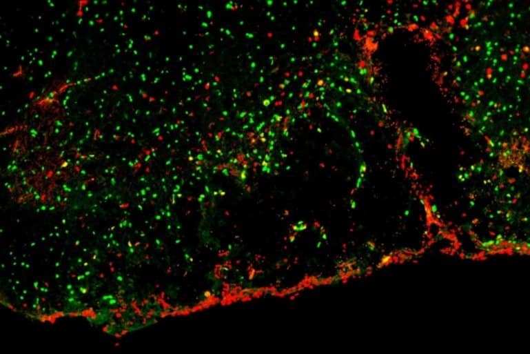
Image: The image is credited to Darpan Saraswat
Original Research: Closed access.
“Heparanome-Mediated Rescue of Oligodendrocyte Progenitor Quiescence following Inflammatory Demyelination” by Darpan Saraswat, R. Ross Welliver, Roopa Ravichandar, Ajai Tripathi, Jessie J. Polanco, Jacqueline Broome, Edward Hurley, Ranjan Dutta, M. Laura Feltri and Fraser J. Sim. Journal of Neuroscience
Abstract
Heparanome-Mediated Rescue of Oligodendrocyte Progenitor Quiescence following Inflammatory Demyelination
The proinflammatory cytokine IFN-γ, which is chronically elevated in multiple sclerosis, induces pathologic quiescence in human oligodendrocyte progenitor cells (OPCs) via upregulation of the transcription factor PRRX1.
In this study using animals of both sexes, we investigated the role of heparan sulfate proteoglycans in the modulation of IFN-γ signaling following demyelination. We found that IFN-γ profoundly impaired OPC proliferation and recruitment following adult spinal cord demyelination. IFN-γ-induced quiescence was mediated by direct signaling in OPCs as conditional genetic ablation of IFNγR1 (Ifngr1) in adult NG2+ OPCs completely abrogated these inhibitory effects. Intriguingly, OPC-specific IFN-γ signaling contributed to failed oligodendrocyte differentiation, which was associated with hyperactive Wnt/Bmp target gene expression in OPCs.
We found that PI-88, a heparan sulfate mimetic, directly antagonized IFN-γ to rescue human OPC proliferation and differentiation in vitro and blocked the IFN-γ-mediated inhibitory effects on OPC recruitment in vivo. Importantly, heparanase modulation by PI-88 or OGT2155 in demyelinated lesions rescued IFN-γ-mediated axonal damage and demyelination. In addition to OPC-specific effects, IFN-γ-augmented lesions were characterized by increased size, reactive astrogliosis, and proinflammatory microglial/macrophage activation along with exacerbated axonal injury and cell death. Heparanase inhibitor treatment rescued many of the negative IFN-γ-induced sequelae suggesting a profound modulation of the lesion environment.
Together, these results suggest that the modulation of the heparanome represents a rational approach to mitigate the negative effects of proinflammatory signaling and rescuing pathologic quiescence in the inflamed and demyelinated human brain.
SIGNIFICANCE STATEMENT The failure of remyelination in multiple sclerosis contributes to neurologic dysfunction and neurodegeneration. The activation and proliferation of oligodendrocyte progenitor cells (OPCs) is a necessary step in the recruitment phase of remyelination.
Here, we show that the proinflammatory cytokine interferon-γ directly acts on OPCs to induce pathologic quiescence and thereby limit recruitment following demyelination. Heparan sulfate is a highly structured sulfated carbohydrate polymer that is present on the cell surface and regulates several aspects of the signaling microenvironment.
We find that pathologic interferon-γ can be blocked by modulation of the heparanome following demyelination using either a heparan mimetic or by treatment with heparanase inhibitor. These studies establish the potential for modulation of heparanome as a regenerative approach in demyelinating disease.