

Summary: According to researchers, bodily responses to pain are controlled by a neural pathway that involves heightened activity in the spinal cord and two parts of the brain-stem.
Source: NIH/NCCIH.
Exposure to uncomfortable sensations elicits a wide range of appropriate and quick reactions, from reflexive withdrawal to more complex feelings and behaviors. To better understand the body’s innate response to harmful activity, researchers at the National Center for Complementary and Integrative Health (NCCIH), part of the National Institutes of Health, have identified activity in the brain that governs these reactions. Using heat as the source of discomfort, experiments conducted by the center’s intramural program showed that bodily responses to pain are controlled by a neural pathway involving heightened activity in the spinal cord and two parts of the brainstem. Results of the study were published in the journal Neuron.
“Much is known about local spinal cord circuits for simple reflexive responses, but the mechanisms underlying more complex behaviors remain poorly understood,” said Alexander T. Chesler, Ph.D., a Stadtman Investigator at NCCIH and senior author of the study. “We set out to describe the brain pathway that controls motor responses and involuntary behaviors when the body is faced with painful experiences.”
Just as people respond to increasingly uncomfortable surfaces like a sandy beach on a hot day by lifting their feet, hopping, and eventually running to a water source, so, too, do laboratory models show a predictable sequence of behaviors. Experiments showed that the parts of a brainstem involved in this circuit are the parabrachial nucleus (PBNI) and the dorsal reticular formation in the medulla (MdD). A specific group of nerve cells in the PBNI is activated by standing on a hot surface, triggering escape responses through connections to the MdD. These PBNI cells express a gene called Tac1, which codes for substances called tachykinins that participate in many functions in the body and contribute to multiple disease processes. The MdD cells involved in this circuit also express Tac1. A different group of cells in the PBNI participates in the aspects of the response to noxious heat that involve the forebrain.
“Our data provide evidence that the PBNI produces streams of information with distinct functional significance,” said Arnab Barik, Ph.D., postdoctoral fellow at NCCIH and one of the study’s authors. “The brainstem-spinal cord pathway identified in this study selectively controls pain response and elicits appropriate behaviors based on sensory input.”
Added Chesler, “This kind of feed-forward circuitry is unique because it is an upward spiral. The more this pathway is activated by harmful activity, the more it reacts, leading to dramatic behavioral responses.”
Knowledge of this brain activity provides new insight into how the body responds to harmful, painful stimuli. The mechanism described in this study can help researchers better understand how pain is encoded in the brain. Further investigation of this circuit may increase understanding of how responses to pain are coordinated with other biological needs, such as feeding and reproduction. These findings may also offer opportunities to understand how the body becomes dysregulated during chronic pain.
This report a basic research finding. Basic research increases our understanding of human behavior and biology, which is foundational to advancing new and better ways to prevent, diagnose, and treat disease. Science is an unpredictable and incremental process — each research advance builds on past discoveries, often in unexpected ways. Most clinical advances would not be possible without the knowledge of fundamental basic research.
Source: NIH/NCCIH
Publisher: Organized by NeuroscienceNews.com.
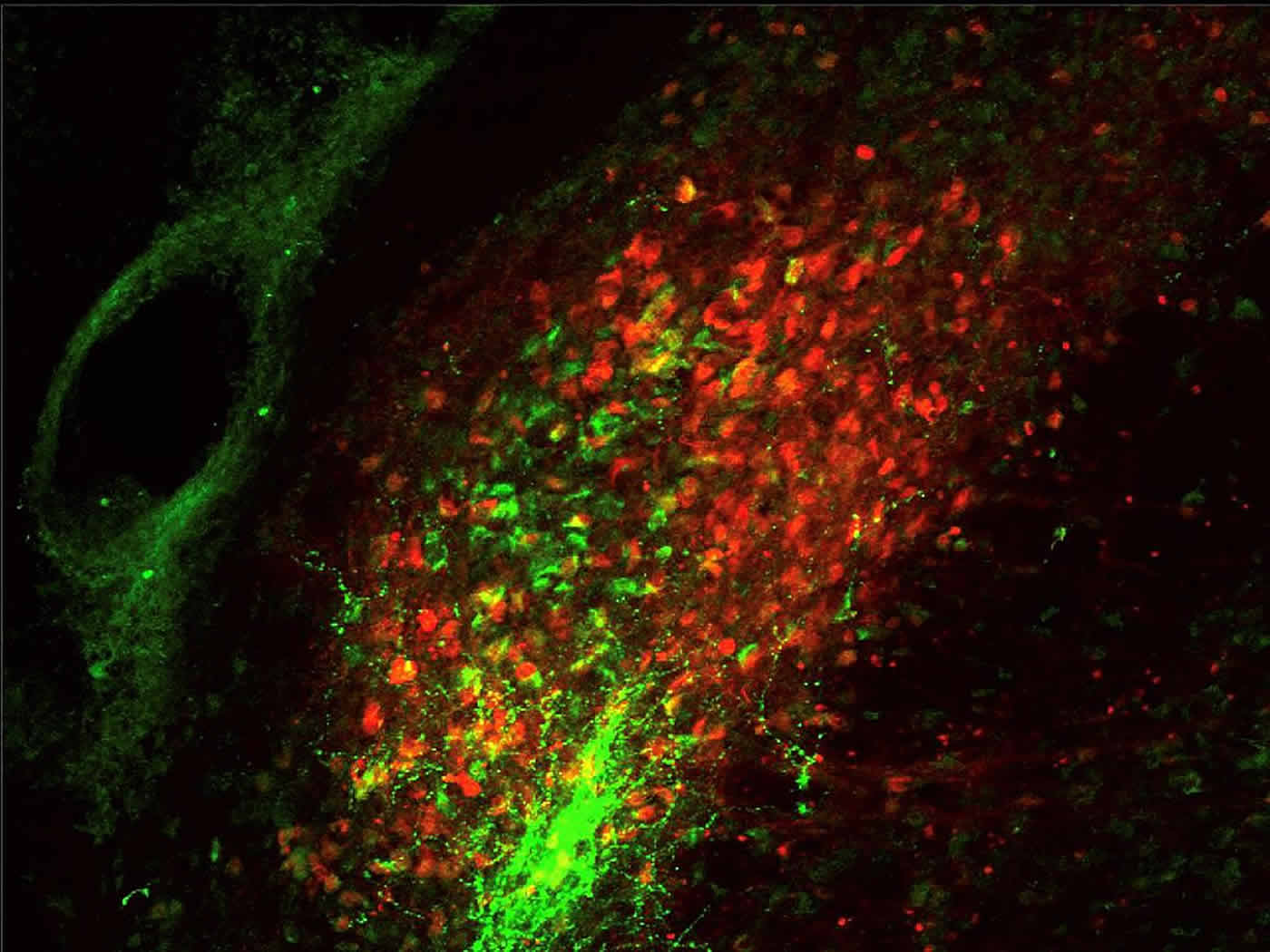
Image Source: NeuroscienceNews.com image is credited to Arnab Barik, Chesler Laboratory, NCCIH.
Original Research: Abstract for “A Brainstem-Spinal Circuit Controlling Nocifensive Behavior” by Arnab Barik, James Hunter Thompson, Mathew Seltzer, Nima Ghitani, and Alexander T. Chesler in Neuron. Published November 15 2019.
doi:10.1016/j.neuron.2018.10.037
[cbtabs][cbtab title=”MLA”]NIH/NCCIH”Study Explains Behavioral Reaction to Painful Experiences.” NeuroscienceNews. NeuroscienceNews, 19 November 2019.
<https://neurosciencenews.com/pain-behavioral-reaction-10221/>.[/cbtab][cbtab title=”APA”]NIH/NCCIH(2019, November 19). Study Explains Behavioral Reaction to Painful Experiences. NeuroscienceNews. Retrieved November 19, 2019 from https://neurosciencenews.com/pain-behavioral-reaction-10221/[/cbtab][cbtab title=”Chicago”]NIH/NCCIH”Study Explains Behavioral Reaction to Painful Experiences.” https://neurosciencenews.com/pain-behavioral-reaction-10221/ (accessed November 19, 2019).[/cbtab][/cbtabs]
Abstract
A Brainstem-Spinal Circuit Controlling Nocifensive Behavior
Response to danger needs to be rapid and appropriate. In humans, nocifensive behaviors often precede conscious pain perception. Much is known about local spinal cord circuits for simple reflexive responses, but the mechanisms underlying more complex behaviors remain poorly understood. We now describe a brainstem circuit that controls escape responses to select noxious stimuli. Tracing experiments characterized a highly interconnected excitatory circuit involving the dorsal spinal cord, parabrachial nucleus (PBNl), and reticular formation (MdD). A combination of chemogenetic, optogenetic, and genetic ablation approaches revealed that PBNl Tac1 neurons are activated by noxious stimuli and trigger robust escape responses to heat through connections to the MdD. Remarkably, MdD Tac1 neurons receive excitatory input from the PBN and target both the spinal cord and PBN; activation of these neurons phenocopies the behavioral effects of PBNl Tac1 neuron stimulation. These findings identify a substrate for controlling appropriate behavioral responses to painful stimuli.


