Summary: Combining virtual reality with neuroimaging, researchers identify how the brain perceives and remembers a novel place or landscape.
Source: NTNU
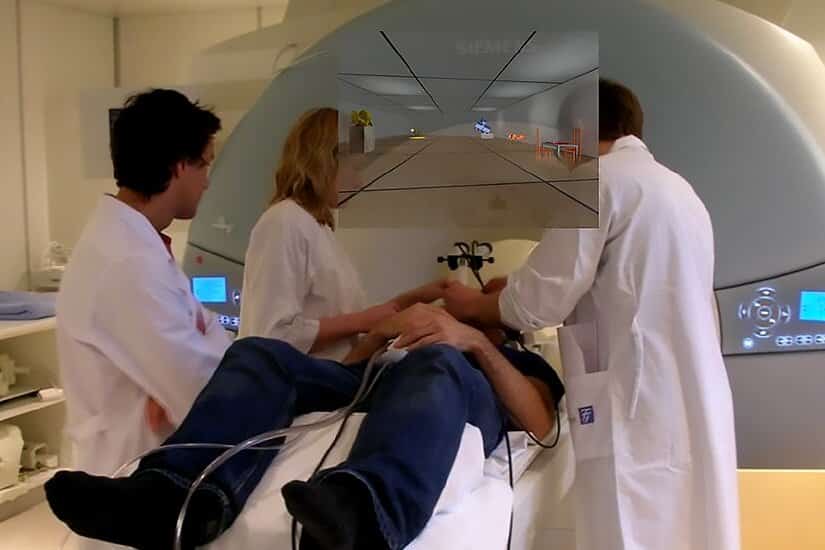
Researchers set up 31 male students to be able to move around various virtual rooms while lying inside an MRI machine that scanned their brains. Equipped with VR glasses and a joystick, participants were given 30 seconds in each room.
The task was to remember the room and the objects. Afterwards, they were allowed to rest, and finally they were given a distracting task where they used other parts of the brain. Brain activity was recorded during all phases.
Researcher Hallvard Røe Evensmoen and professor Asta Kristine Håberg at NTNU’s Department of Neuromedicine and Movement Science used functional MRI and self-developed computer systems in the research project.
The systems provided new knowledge about how the brain perceives and remembers a new place or landscape.
The research report was published in the renowned scientific journal Cell Reports.
Surprising discovery #1: We remember where, but not what
It turned out that the subjects had an easier time remembering where things were placed than exactly what things were placed in each location.
“In the most extreme cases, the subject could remember the position pattern of the landmarks exactly, without remembering which landmarks were located where. Our brain is pretty set on remembering the positions of landmarks,” says Evensmoen.
The analyses of the data from the MRI scan showed that remembering positions and remembering objects associated with the positions are represented in two different but synchronized networks in the brain.
Previous research has made no distinction between these two parts of memory.
“The coding of the functions is different, but they’re located in the same area of the brain, the medial part of the temporal lobe, where the hippocampus and other memory areas are located,” says Håberg.
Surprising discovery 2: Good networks mean everything
Their research also revealed that more parts of the brain than previously thought are involved in remembering a new landscape.
An extensive network of brain areas has to work closely together when we form a cognitive map—an idea in our brain—of a new landscape.
“The cognitive map is formed in a network of brain areas with synchronized activation and overlapping functions,” say the researchers.
Areas that are important for sight and feelings are also important for remembering a new place.
It has been common to assume that spatial memory is organized with the hippocampus as a kind of central hub, but this is not entirely true according to Evensmoen’s and Håberg’s latest research.
The subjects with the most parallel connections in the brain were the ones who were best at remembering exactly where in the environment objects were located.
“Effective networks are crucial for how well you remember positions and objects in a new place,” Evensmoen says.
Surprising discovery 3: The brain worked mostly during rest
The experiment also showed that brain activity increased and that even more connections in the brain were activated during the rest period after participants had finished moving around each room.

“A secondary finding in our research was that the participants’ brain activity was greater when they were resting than when they moved around the rooms. The brain continued to work on establishing a cognitive map even though they no longer were moving around in the environment,” say Evensmoen and Håberg. They say this a very interesting discovery and that it fits well with previous knowledge.
“What you experience during the day continues to consolidate in the brain when you sleep. Taking a nap in the middle of the day strengthens memory,” Håberg adds.
Plan to continue memory studies
“What will be useful now is that we have a new starting point for studying memory with the results and methods we’ve developed. We’ll be able to see if there are differences in other types of memory than spatial, like illness or aging, and whether these principles also apply to them,” says Håberg.
More precise knowledge can make it easier to detect early signs of disease versus normal aging in the elderly, for example. It can also provide a basis for better exercise programs for people who have various forms of brain damage.
About this memory research news
Author: Wenche Margrethe Kulmo
Source: NTNU
Contact: Wenche Margrethe Kulmo – NTNU
Image: The image is credited to NTNU
Original Research: Open access.
“Allocentric representation in the human amygdala and ventral visual stream” by Hallvard Røe Evensmoen et al. Cell Reports
Abstract
Allocentric representation in the human amygdala and ventral visual stream
Highlights
- •The allocentric medial temporal lobe network is globally efficient, with no main hub
- •Ventral visual stream plays a particularly central role in the allocentric network
- •Allocentric representations are in distinct but neighboring neural populations
- •The human brain encodes both scaled and true Euclidean representations
Summary
The hippocampus and the entorhinal cortex are considered the main brain structures for allocentric representation of the external environment.
Here, we show that the amygdala and the ventral visual stream are involved in allocentric representation.
Thirty-one young men explored 35 virtual environments during high-resolution functional magnetic resonance imaging (fMRI) of the medial temporal lobe (MTL) and were subsequently tested on recall of the allocentric pattern of the objects in each environment—in other words, the positions of the objects relative to each other and to the outer perimeter.
We find increasingly unique brain activation patterns associated with increasing allocentric accuracy in distinct neural populations in the perirhinal cortex, parahippocampal cortex, fusiform cortex, amygdala, hippocampus, and entorhinal cortex.
In contrast to the traditional view of a hierarchical MTL network with the hippocampus at the top, we demonstrate, using recently developed graph analyses, a hierarchical allocentric MTL network without a main connector hub.


