

Summary: Researchers use fast fMRI to track the brain activity that occurs during thought. The study could be a significant step toward mapping the neural networks responsible for attention, perception and other cognitive functions.
Source: NIH/NIBIB.
Fast fMRI tracks brain activity during human thought for first time.
By significantly increasing the speed of functional MRI (fMRI), researchers funded by the National Institute of Biomedical Imaging and Bioengineering (NIBIB) have been able to image rapidly fluctuating brain activity during human thought. fMRI measures changes in blood oxygenation, which were previously thought to be too slow to detect the subtle neuronal activity associated with higher order brain functions. The new discovery that fast fMRI can detect rapid brain oscillations is a significant step towards realizing a central goal of neuroscience research: mapping the brain networks responsible for human cognitive functions such as perception, attention, and awareness.
“A critical aim of the President’s BRAIN Initiative is to move neuroscience into a new realm where we can identify and track functioning neural networks non-invasively,” explains Guoying Liu, Ph.D., Director of the NIBIB program in Magnetic Resonance Imaging. “This work demonstrates the potential of fMRI for mapping healthy neural networks as well as those that may contribute to neurological diseases such as dementia and other mental health disorders, which are significant national and global health problems.”
fMRI works by detecting local increases in oxygen as blood is delivered to a working part of the brain. The technique has been instrumental for identifying which areas in the brain control functions such as vision, hearing, or touch. However, standard fMRI can only detect the blood flow coming to replenish an area of the brain several seconds after it has performed a function. It was generally accepted that this was the limit of what could be detected by fMRI—identification of a region in the brain that had responded to a large stimulus, such as a continuous 30 second “blast” of bright light.
Combining several new techniques, Jonathan R. Polimeni, Ph.D., senior author of the study, and his colleagues at Harvard’s Athinoula A. Martinos Center for Biomedical Imaging, applied fast fMRI in an effort to track neuronal networks that control human thought processes, and found that they could now measure rapidly oscillating brain activity. The results of this groundbreaking work are reported in the October 2016 issue of the Proceedings of the National Academy of Sciences.
The researchers used fast fMRI in human volunteers observing a rapidly fluctuating checkerboard pattern. The fast fMRI was able to detect the subtle and very rapid oscillations in cerebral blood flow in the brain’s visual cortex as the volunteers observed the changing pattern.
“The oscillating checkerboard pattern is a more “naturalistic” stimulus, in that its timing is similar to the very subtle neural oscillations made during normal thought processes,” explains Polimeni. “The fast fMRI detects the induced neural oscillations that allow the brain to understand what the eye is observing — the changing checkerboard pattern. These subtle oscillations were completely undetectable with standard fMRI. This exciting result opens the possibility of using fast fMRI to image neural networks as they guide the process of human thought.”
One such possibility is suggested by first author of the study Laura D. Lewis, Ph.D. “This technique now gives us a method for obtaining much more detailed information about the complex brain activity that takes place during sleep, as well as other dynamic switches in brain states, such as when under anesthesia and during hallucinations.”
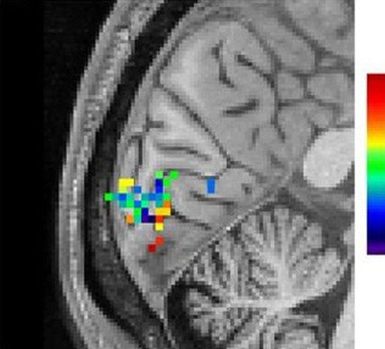
fMRI signals in the brain’s visual cortex as an individual views the checkerboard stimulus. The green, blue and dark blue spots represent signals produced within a fraction of a second after stimulation with dark blue being the fastest responding areas. NeuroscienceNews.com image is credited to Lewis, et al. PNAS. Oct. 2016.
Concludes Polimeni, “It had always been thought that fMRI had the potential to play a major role in these types of studies. Meaningful progress in cognitive neuroscience depends on mapping patterns of brain activity, which are constantly and rapidly changing with every experience we have. Thus, we are extremely excited to see our work contribute significantly to achieving this goal.”
The results of this work contributed to Dr. Polimeni’s recent grant award from the NIH BRAIN Initiative. The BRAIN funding will be used to extend studies using fast fMRI to continue improvement in the fine scale mapping of human brain function.
Funding: The work was funded by the National Institutes of Health (NIH) through grants EB011498, EB019437, and EB015896 from the National Institute for Biomedical Imaging and Bioengineering. Additional funding was provided by several NIH Shared Resource Instrumentation Grants, and the Athinoula A. Martinos Center for Biomedical Imaging.
Source: Thomas Johnson – NIH/NIBIB
Image Source: NeuroscienceNews.com image is credited to Lewis, et al. PNAS. Oct. 2016.
Original Research: Full open access research for “Fast fMRI can detect oscillatory neural activity in humans” by Laura D. Lewis, Kawin Setsompop, Bruce R. Rosen, and Jonathan R. Polimeni in PNAS. Published online October 11 2016 doi:10.1073/pnas.1608117113
[cbtabs][cbtab title=”MLA”]NIH/NIBIB “Imaging Technique Can See You Think.” NeuroscienceNews. NeuroscienceNews, 1 December 2016.
<https://neurosciencenews.com/neuroscience-thinking-neuroimaging-5640/>.[/cbtab][cbtab title=”APA”]NIH/NIBIB (2016, December 1). Imaging Technique Can See You Think. NeuroscienceNew. Retrieved December 1, 2016 from https://neurosciencenews.com/neuroscience-thinking-neuroimaging-5640/[/cbtab][cbtab title=”Chicago”]NIH/NIBIB “Imaging Technique Can See You Think.” https://neurosciencenews.com/neuroscience-thinking-neuroimaging-5640/ (accessed December 1, 2016).[/cbtab][/cbtabs]
Abstract
Fast fMRI can detect oscillatory neural activity in humans
Oscillatory neural dynamics play an important role in the coordination of large-scale brain networks. High-level cognitive processes depend on dynamics evolving over hundreds of milliseconds, so measuring neural activity in this frequency range is important for cognitive neuroscience. However, current noninvasive neuroimaging methods are not able to precisely localize oscillatory neural activity above 0.2 Hz. Electroencephalography and magnetoencephalography have limited spatial resolution, whereas fMRI has limited temporal resolution because it measures vascular responses rather than directly recording neural activity. We hypothesized that the recent development of fast fMRI techniques, combined with the extra sensitivity afforded by ultra-high-field systems, could enable precise localization of neural oscillations. We tested whether fMRI can detect neural oscillations using human visual cortex as a model system. We detected small oscillatory fMRI signals in response to stimuli oscillating at up to 0.75 Hz within single scan sessions, and these responses were an order of magnitude larger than predicted by canonical linear models. Simultaneous EEG–fMRI and simulations based on a biophysical model of the hemodynamic response to neuronal activity suggested that the blood oxygen level-dependent response becomes faster for rapidly varying stimuli, enabling the detection of higher frequencies than expected. Accounting for phase delays across voxels further improved detection, demonstrating that identifying vascular delays will be of increasing importance with higher-frequency activity. These results challenge the assumption that the hemodynamic response is slow, and demonstrate that fMRI has the potential to map neural oscillations directly throughout the brain.
“Fast fMRI can detect oscillatory neural activity in humans” by Laura D. Lewis, Kawin Setsompop, Bruce R. Rosen, and Jonathan R. Polimeni in PNAS. Published online October 11 2016 doi:10.1073/pnas.1608117113


