Summary: New research uncovers the exact immune-to-brain pathway that drives the loss of social motivation during sickness. Scientists showed that when the cytokine IL-1β binds to receptors on neurons within the dorsal raphe nucleus, it activates a circuit that reduces social interaction.
This pathway acts independently of lethargy, revealing that social withdrawal is an actively generated behavioral response rather than a passive one. The findings highlight a precise neural mechanism by which the immune system reshapes behavior during illness.
Key Facts:
- Immune–Brain Link: IL-1β directly activates neurons in the dorsal raphe nucleus to suppress social behavior.
- Specific Circuit: Only the pathway from the DRN to the lateral septum reproduced full social withdrawal.
- Not Just Fatigue: Blocking this circuit prevented withdrawal but did not stop illness-related lethargy.
Source: Picower Institute at MIT
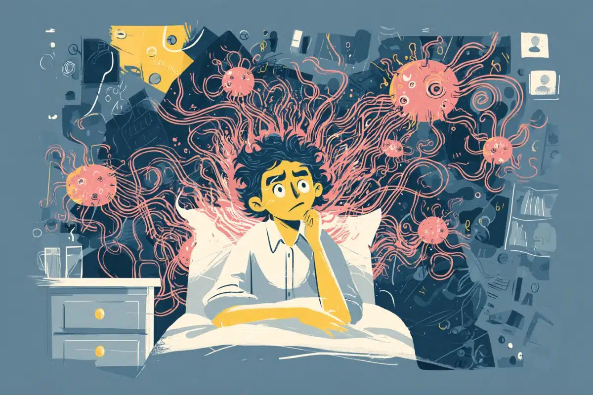
“I just can’t make it tonight. You have fun without me.” Across much of the animal kingdom, when infection strikes, social contact shuts down.
A new study details how the immune and central nervous systems implement this sickness behavior.
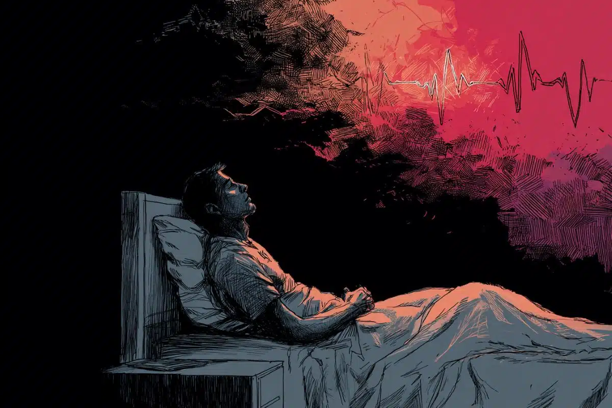
It makes perfect sense that when we’re battling an infection, we lose our desire to be around others. That protects them from getting sick and lets us get much needed rest. What hasn’t been as clear is how this behavior change happens.
In the research published Nov. 25 in Cell, scientists at The Picower Institute for Learning and Memory of MIT and collaborators used multiple methods to demonstrate causally that when the immune system cytokine interleukin-1 beta (IL-1β) reaches the IL-1 receptor 1 (IL-1R1) on neurons in a brain region called the dorsal raphe nucleus, that activates connections with the intermediate lateral septum to shut down social behavior.
“Our findings show that social isolation following immune challenge is self-imposed and driven by an active neural process, rather than a secondary consequence of physiological symptoms of sickness, such as lethargy,” said study co-senior author Gloria Choi, associate professor in The Picower Institute and MIT’s Department of Brain and Cognitive Sciences.
Jun Huh, Harvard Medical School associate professor of immunology, is the paper’s co-senior author. The lead author is Liu Yang, a research scientist in Choi’s lab.
A molecule and its receptor
Choi and Huh’s long collaboration have identified other cytokines that affect social behavior by latching on to their receptors in the brain, so in this study their team hypothesized that the same kind of dynamic might cause social withdrawal during infection. But which cytokine? And what brain circuits might be affected?
To get started, Yang and her colleagues injected 21 different cytokines into the brains of mice, one by one, to see if any triggered social withdrawal the same way that giving mice LPS (a standard way of simulating infection) did. Only IL-1β injection fully recapitulated the same social withdrawal behavior as LPS. That said, IL-1β also made the mice more sluggish.
IL-1β affects cells when it hooks up with the IL-1R1, so the team next went looking across the brain for where the receptor is expressed. They identified several regions and examined individual neurons in each.
The dorsal raphe nucleus (DRN) stood out among regions, both because it is known to modulate social behavior and because it is situated next to the cerebral aqueduct, which would give it plenty of exposure to incoming cytokines in cerebrospinal fluid.
The experiments identified populations of DRN neurons that express the IL-1R1, including many involved in making the crucial neuromodulatory chemical serotonin.
From there, Yang and the team demonstrated that IL-1β activates those neurons, and that activating the neurons promotes social withdrawal. Moreover, they showed that inhibiting that neural activity prevented social withdrawal in mice treated with IL-1β, and they showed that shutting down the IL-1R1 in the DRN neurons also prevented social withdrawal behavior after IL-1β injection or LPS exposure.
Notably, these experiments did not change the lethargy that followed IL-1β or LPS, helping to demonstrate that social withdrawal and lethargy occur through different means.
“Our findings implicate IL-1β as a primary effector driving social withdrawal during systemic immune activation,” the researchers wrote in Cell.
Tracing the circuit
With the DRN identified as the site where neurons receiving IL-1β drove social withdrawal, the next question was what circuit they effected that behavior change through. The team traced where the neurons make their circuit projections and found several regions that have a known role in social behavior.
Using optogenetics, a technology that engineers cells to become controllable with flashes of light, the scientists were able to activate the DRN neurons’ connections with each downstream region. Only activating the DRN’s connections with the intermediate lateral septum caused the social withdrawal behaviors seen with IL-1β injection or LPS exposure.
In a final test, they replicated their results by exposing some mice to salmonella.
“Collectively, these results reveal a role for IL-1R1-expressing DRN neurons in mediating social withdrawal in response to IL-1β during systemic immune challenge,” the researchers wrote.
Though the study revealed the cytokine, neurons and circuit responsible for social withdrawal in mice in detail and with demonstrations of causality, the results still inspire new questions. One is whether IL-1R1 neurons affect other sickness behaviors. Another is whether serotonin has a role in social withdrawal or other sickness behaviors.
In addition to Yang, Choi and Huh, the paper’s other authors are Matias Andina, Mario Witkowski, Hunter King, and Ian Wickersham.
Funding: Funding for the research came from The National Institute of Mental Health, the National Research Foundation of Korea, the Denis A. and Eugene W. Chinery Fund for Neurodevelopmental Research, the Jeongho Kim Neurodevelopmental Research Fund, Perry Ha, the Simons Center for the Social Brain, the Simons Foundation Autism Research Initiative, The Picower Institute for Learning and Memory, and The Freedom Together Foundation.
Key Questions Answered:
A: An immune molecule called IL-1β activates a specific brain receptor that shuts down social behavior.
A: Neurons in the dorsal raphe nucleus activate pathways to the lateral septum to suppress social interaction.
A: No — the study shows it is an active, separate neural process, not a side-effect of feeling sluggish.
Editorial Notes:
- This article was edited by a Neuroscience News editor.
- Journal paper reviewed in full.
- Additional context added by our staff.
About this neuroscience and social isolation research news
Author: David Orenstein
Source: Picower Institute at MIT
Contact: David Orenstein – Picower Institute at MIT
Image: The image is credited to Neuroscience News
Original Research: Open access.
“IL-1R-positive dorsal raphe neurons drive self-imposed social withdrawal in sickness” by Gloria Choi et al. Cell
Abstract
IL-1R-positive dorsal raphe neurons drive self-imposed social withdrawal in sickness
Sick animals exhibit behavioral changes that extend beyond physiological symptoms, such as appetite loss and hypoactivity, and include a decline in social interactions.
While social isolation during sickness has been recognized to have the evolutionary benefit of staving off disease spread, the molecular and neural mechanisms underlying this response remain unclear.
Cytokines—immune-derived signaling molecules—have emerged as neuromodulators impacting brain function during inflammation.
Through behavioral screening, we identify a unique role for the cytokine interleukin-1β (IL-1β) in promoting social withdrawal during sickness. IL-1β directly modulates the activity of IL-1R1-expressing neurons in the dorsal raphe nucleus (DRN) (IL-1R1DRN).
Activation of these neurons is sufficient to elicit social withdrawal, while their inhibition or genetic deletion of IL-1R1 rescues self-imposed social isolation during systemic inflammation.
Our findings reveal a neural mechanism that actively promotes social disengagement in sick animals, highlighting the role of IL-1R1DRN neurons in driving these behavioral adaptations.