Summary: A groundbreaking study reveals that neurons can burn fat, challenging the long-held belief that the brain relies solely on glucose for fuel. Researchers discovered that when glucose is scarce, synaptic activity triggers neurons to break down lipid droplets and use the resulting fatty acids for energy.
This lipid metabolism process is regulated by electrical activity and is essential for maintaining brain function, especially under stress. Blocking this fat-burning pathway in mice caused a dramatic drop in body temperature, revealing just how crucial it is for survival.
Key Facts:
- Fat as Fuel: Synaptic activity enables neurons to break down fat droplets for energy, particularly when glucose is scarce.
- Genetic Clue: The DDHD2 gene plays a key role in this process; mutations in it are linked to neurological disorders.
- Functional Evidence: Blocking fat-burning enzymes in mice induced torpor, highlighting the importance of lipid metabolism in the brain.
Source: Weill Cornell University
While glucose, or sugar, is a well-known fuel for the brain, Weill Cornell Medicine researchers have demonstrated that electrical activity in synapses—the junctions between neurons where communication occurs—can lead to the use of lipid or fat droplets as an energy source.
The study, published July 1 in Nature Metabolism, challenges “the long-standing dogma that the brain doesn’t burn fat,” said principal investigator Dr. Timothy A. Ryan, professor of biochemistry and of biochemistry in anesthesiology, and the Tri-Institutional Professor in the Department of Biochemistry at Weill Cornell Medicine.

The paper’s lead author, Dr. Mukesh Kumar, a postdoctoral associate in biochemistry at Weill Cornell Medicine who has been studying the cell biology of fat droplets, suggested that it makes sense that fat may play a role as an energy source in the brain like it does with other metabolically demanding tissues, such as muscle.
The research team was particularly intrigued by the DDHD2 gene, which encodes a lipase, or enzyme that helps break down fat. Mutations in DDHD2 are linked to a type of hereditary spastic paraplegia, a neurological condition that causes progressive stiffness and weakness in the legs, in addition to cognitive deficits.
Prior research by other investigators has demonstrated that blocking this enzyme in mice causes a build-up of triglycerides—or fat droplets that store energy—throughout the brain. “To me, this was evidence that maybe the reason we claim the brain doesn’t burn fat is because we never see the fat stores,” Dr. Ryan said.
Research Demonstrates Lipids Have an Important Role
The current study explored whether the lipid droplets that build up in the absence of DDHD2 are used as fuel by the brain, particularly when glucose isn’t present, Dr. Ryan said.
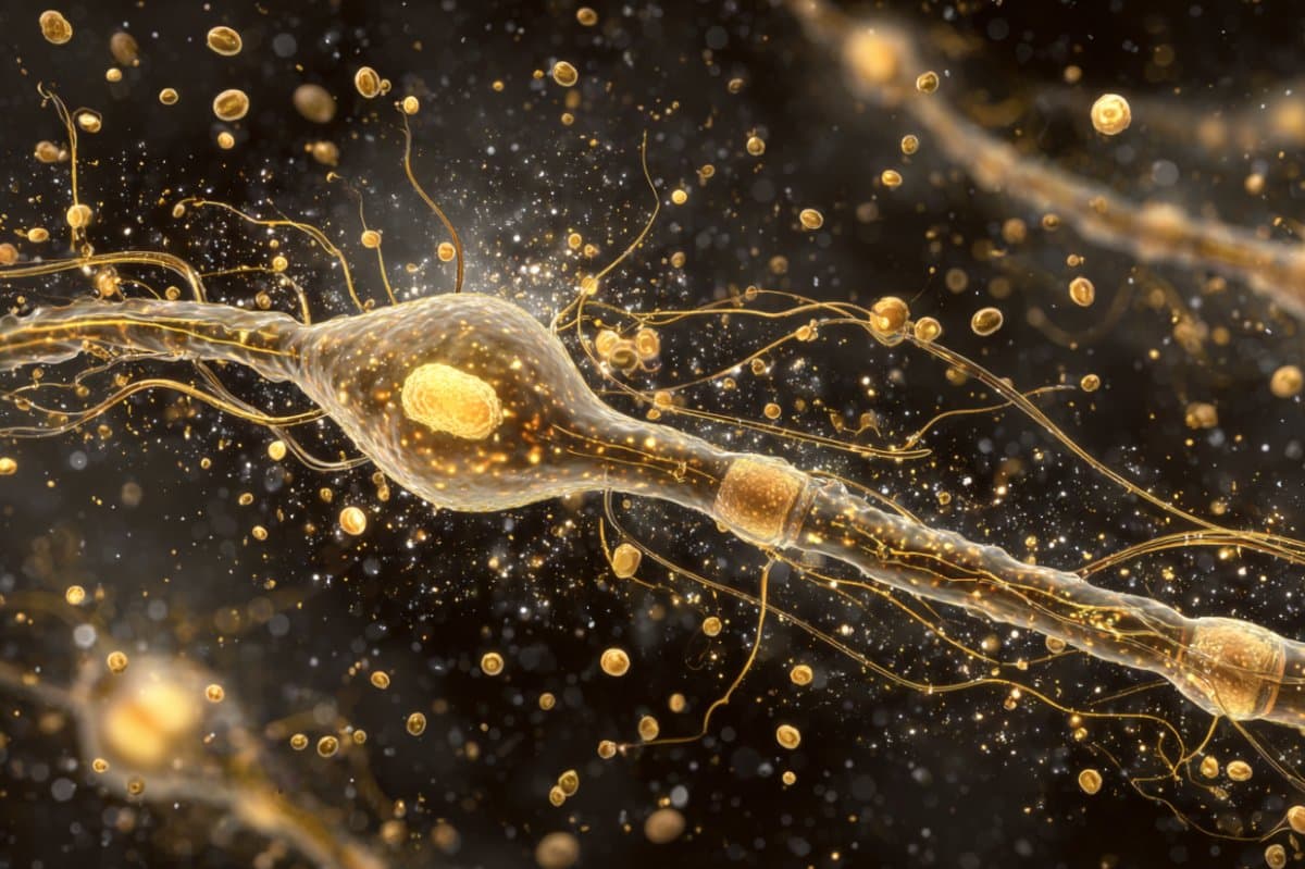
Dr. Kumar found that when a synapse contains a lipid droplet filled with triglycerides in mice without DDHD2, neurons can break down this fat into fatty acids and send it to the mitochondria—the cell’s energy factories—so they can produce adenosine triphosphate (ATP), the energy the cell needs to function.
“The process of being able to use the fat is controlled by the electrical activity of the neurons, and I was shocked by this finding,” Dr. Ryan said. “If the neuron is busy, it drives this consumption. If it’s at rest, the process isn’t happening.”
In another study, researchers injected a small molecule into mice to block the enzyme carnitine palmitoyltransferase 1 (CPT1), which helps transport fatty acids into the mitochondria for energy production.
Blocking CPT1 prevented the brain from using fat droplets, which then led to torpor, a hibernation-like state, in which the body temperature rapidly plummets and the heartbeat slows.
“This response convinced us that that there’s an ongoing need for the brain to use these lipid droplets,” Dr. Ryan said.
Implications for Future Research
This research may encourage the further investigation of neurodegenerative conditions and the role of lipids in the brain. Glucose fluctuations or low levels of glucose can occur with aging or neurological disease, but fatty acids broken down from lipid droplets may help to maintain the brain’s energy, Dr. Kumar said.
“We don’t know where this research will go in terms of neurodegenerative conditions, but some evidence suggests that accumulation of fat droplets in the neurons may occur in Parkinson’s disease,” he said.
Researchers also need to better understand the interplay between glucose and lipids in the brain, Dr. Ryan said.
“By learning more about these molecular details, we hope to ultimately unlock explanations for neurodegeneration, which would give us opportunities for finding ways to protect the brain.”
About this metabolism and neuroscience research news
Author: Corinne Esposito
Source: Weill Cornell University
Contact: Corinne Esposito – Weill Cornell University
Image: The image is credited to Neuroscience News
Original Research: Open access.
“Triglycerides are an important fuel reserve for synapse function in the brain” by Timothy A. Ryan et al. Nature Metabolism
Abstract
Triglycerides are an important fuel reserve for synapse function in the brain
Proper fuelling of the brain is critical to sustain cognitive function, but the role of fatty acid (FA) combustion in this process has been elusive.
Here we show that acute block of a neuron-specific triglyceride lipase, DDHD2 (a genetic driver of complex hereditary spastic paraplegia), or of the mitochondrial lipid transporter CPT1 leads to rapid onset of torpor in adult male mice.
These data indicate that in vivo neurons are probably constantly fluxing FAs derived from lipid droplets (LDs) through β-oxidation to support neuronal bioenergetics.
We show that in dissociated neurons, electrical silencing or blocking of DDHD2 leads to accumulation of neuronal LDs, including at nerve terminals, and that FAs derived from axonal LDs enter mitochondria in an activity-dependent fashion to drive local mitochondrial ATP production.
These data demonstrate that nerve terminals can make use of LDs during electrical activity to provide metabolic support and probably have a critical role in supporting neuron function in vivo.


