Summary: A group of hippocampal neurons show rhythmic activity at different frequencies in the desynchronized state, but can align their rhythmic frequency to produce a synchronized brain rhythm upon activation.
Source: Institute for Experimental Medicine
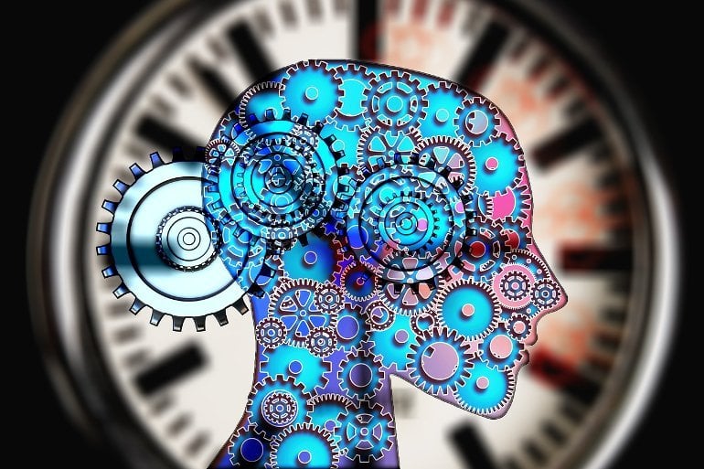
In the 17th century, the Dutch scientist Christiaan Huygens hung two of his recently invented pendulum clocks on a wooden beam and observed that as time passed, the clocks aligned their beats. He reported this finding, which he called an “odd sympathy,” in 1665. Three and a half centuries later, neurons in the brain were found to sync their activities in a similar way.
Neurons in the brain often synchronize in quasi-rhythmic activities, collectively generating “brain waves” sometimes detectable even from outside the skull using electroencephalography. Synchronizing in such rhythms helps neurons to exchange information efficiently, which is crucial to perform important functions like learning, memory, attention, perception, and motion.
How these rhythms are generated, maintained, and abolished to fit ever-changing needs for seamless operation of the brain is an area of active research.
In a new study published today in Cell Reports, a team of neuroscientists led by Principal Investigator Balázs Hangya MD Ph.D. at the Institute of Experimental Medicine, Budapest, Hungary found that a group of neurons in the brain show rhythmic activity at different frequencies in the desynchronized state, like individual clocks, but they can align their rhythmic frequencies to produce a synchronized brain rhythm upon activation, similar to the pendulum clocks in Huygens’ experiment.
To probe the brain’s sync mechanisms, the team recorded special neurons of a deep brain structure called the medial septum. These neurons form a “pacemaker network,” which generates the 4-12 Hz “theta” rhythm in a structure called the hippocampus, responsible for encoding episodic memory traces of the events we experience. It is well established that the hippocampal theta oscillation is important for memory, but the exact mechanism by which it is generated is not well understood.

“To understand how cells in the medial septum synchronize, one should record the activity of several cells at once. In addition to this multichannel deep brain recording, a parallel recording of hippocampal activity is also necessary to understand the output signal of the medial septum. This is still technically challenging, so these recordings can only be performed by skilled experimenters,” said Hangya.
To tackle this challenge, Hangya’s lab teamed up with other groups led by professors István Ulbert, Viktor Varga and Szabolcs Káli to probe the medial septal “pacemaker network” in both rats and mice, awake or under anesthesia. The Huygens synchronization mechanism was present under all tested conditions and could also be reproduced by a detailed computational model of the medial septal “pacemaker network.”
The authors speculate that Huygens synchronization can be a general synchronization mechanism across brain circuits in different species, including humans.
“I think we have come up with a new idea about the origin of network synchronization. The fact that medial septal inhibitory cells synchronize their frequencies upon activation by local excitatory inputs was previously unknown,” says Barnabás Kocsis, the first author of the article.
The brain’s sync mechanisms can go wrong in disease, leading to problems with memory and attention, and even contributing to serious conditions like schizophrenia, epilepsy and Alzheimer’s disease.
The researchers hope that a better understanding of how brain networks synchronize may eventually lead us to improved therapies of these illnesses.
About this neuroscience research news
Author: Press Office
Source: Institute of Experimental Medicine
Contact: Press Office – Institute of Experimental Medicine
Image: The image is in the public domain
Original Research: The findings will appear in Current Biology


