Source: Using an advanced microscopy technique, researchers discovered the role adhesion molecules play in guiding neurons into their correct location.
Source: Yale
In a new study, Yale researchers used an advanced microscopy technique that allowed them to follow a single neuron in the embryo of a worm as it found its neurological home.
The research team, led by Titas Sengupta, working in the lab of Daniel Colón-Ramos, the Dorys McConnell Duberg Professor of Neuroscience and Cell Biology, found that adhesion molecules grab onto neurons which then are essentially pulled into a specific layer or neighborhood and are “zippered” into place.
The findings were published in the journal eLife.
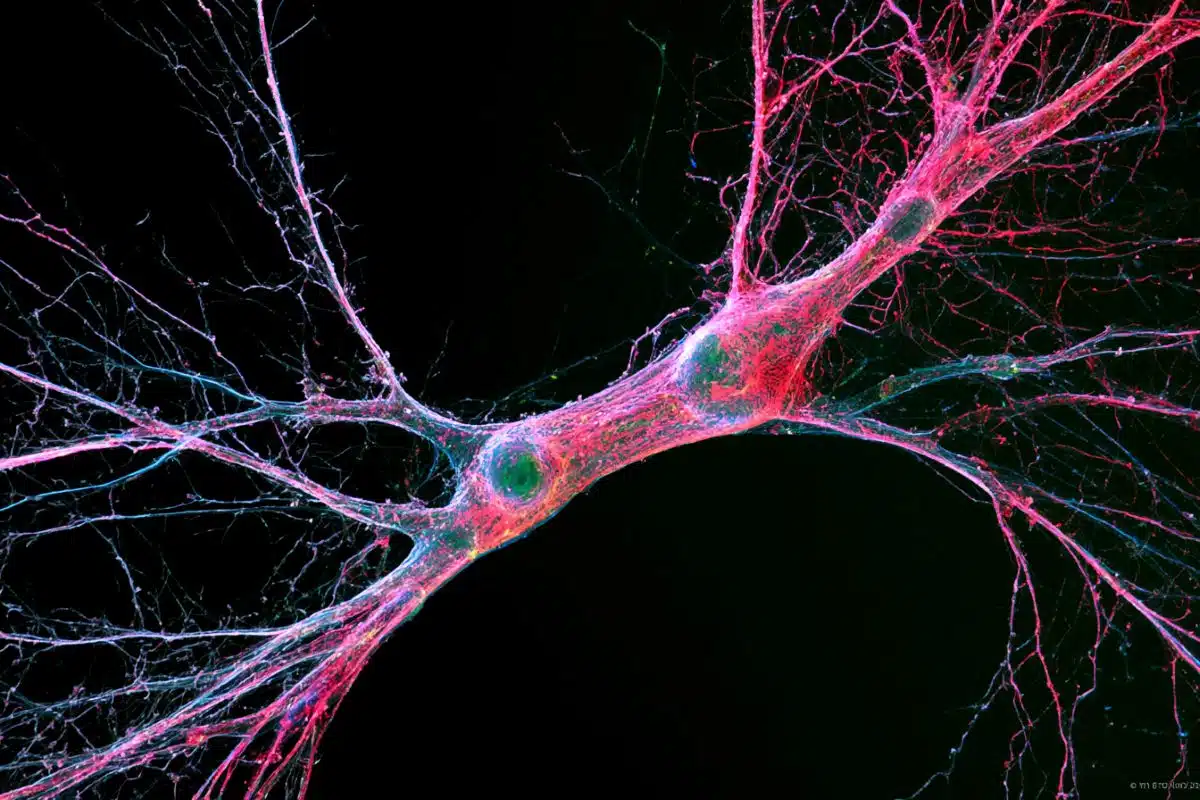
AIB single neurite is placed along two distinct neighborhoods in the nerve ring. (A) Schematic of an adult/larval C. elegans showing an AIB neuron (cyan) and its posterior (orange) and anterior (magenta) neighborhoods in the head. The AIB neurite has a proximal neurite segment (orange arrow), a posterior-anterior shift at the dorsal midline (dashed line) and a distal neurite segment (magenta arrow; on the other side of the worm, behind the pharynx, which is in gray). The neon-colored outline represents the nerve ring neuropil. The terms ‘proximal’ or ‘distal’ neurite segments refer to the relationship of the neurite segment to the AIB cell body. The neighborhoods in which the ‘proximal’ and ‘distal’ neurite segments are positioned are referred to as the ‘posterior’ or ‘anterior’ neighborhoods, respectively, because of their position along the anterior-posterior axis of the worm. Note that this schematic only shows one neuron of the AIB pair. Cell body is marked with an asterisk. (B) Magnified schematic of AIB and its neighborhoods in (A, C) Representative confocal image showing the lateral view of an AIB neuron labeled with cytoplasmic mCherry (cyan). (D) Representative confocal image showing an AIB neuron labeled with cytoplasmic mCherry (cyan); and RIM motor neuron of the anterior neighborhood labeled with cytoplasmic GFP (magenta) in lateral view. Note the colocalization of the AIB distal neurite (but not the proximal neurite) with the anterior neighborhood marker RIM (compare with E). (E) As (D), but with AIB (cyan) and AWC and ASE sensory neurons of the posterior neighborhood (orange). Note the colocalization of the AIB proximal neurite (but not the distal neurite) with the posterior neighborhood markers AWC and ASE (compare with D). (F–J) Same as A–E but in axial view indicated by the arrow in (F). The worm head is tilted in this view to make the two neurite segments in the two neighborhoods visible. Note shift in H (arrows), corresponding to AIB neurite shifting neighborhoods (compare I and J). (K,L) Volumetric reconstruction from the JSH electron microscopy connectome dataset (White et al., 1986) of AIBL (K), and AIBL overlaid on nerve ring strata (L), in lateral view, with S2 and S3 strata (named as in Moyle et al., 2021), containing anterior and posterior neighborhoods, respectively. (M) Volumetric reconstruction of AIBL and AIBR in axial view (from the JSH dataset White et al., 1986). Note the shift in neighborhoods by AIBL and AIBR, at the dorsal midline (dashed line), forms a chiasm (also see Figure 1—figure supplement 1). (N) Schematic of M highlighting the AIB neighborhoods for context and the dorsal midline with a dashed line (AIB neighborhoods, synaptic polarity and resulting network properties also shown in Figure 1—figure supplement 2). Scale bar = 10 μm for A–J and 3 μm for K–N. Credit: The researchers
In a video, Colón-Ramos describes what the technology revealed.
“What we’re able to do with this microscope is track those events as they’re actually occurring and ascribe specific genetic functions to these molecules that we’re identifying in that sequence of events,” he said.
Credit: Yale
These insights, he said, will help scientists determine the molecular roots of many early developmental diseases.
About this neuroscience research news
Author: Press Office Source: Yale Contact: Press Office – Yale Image: The image is credited to the researchers
Differential adhesion regulates neurite placement via a retrograde zippering mechanism
During development, neurites and synapses segregate into specific neighborhoods or layers within nerve bundles.
The developmental programs guiding placement of neurites in specific layers, and hence their incorporation into specific circuits, are not well understood. We implement novel imaging methods and quantitative models to document the embryonic development of the C. elegans brain neuropil, and discover that differential adhesion mechanisms control precise placement of single neurites onto specific layers.
Differential adhesion is orchestrated via developmentally regulated expression of the IgCAM SYG-1, and its partner ligand SYG-2. Changes in SYG-1 expression across neuropil layers result in changes in adhesive forces, which sort SYG-2-expressing neurons. Sorting to layers occurs, not via outgrowth from the neurite tip, but via an alternate mechanism of retrograde zippering, involving interactions between neurite shafts.
Our study indicates that biophysical principles from differential adhesion govern neurite placement and synaptic specificity in vivo in developing neuropil bundles.