

Summary: vDISCO, a new imaging technique based on a method for making tissue and organs transparent, is helping researchers to gain unprecedented insights into neuron interactions in the central nervous system.
Source: LMU.
A new imaging technique developed by LMU researchers is garnering lots of attention. Based on a method for making tissues, organs and even whole organisms transparent, it promises to transform studies of the nervous system in particular.
A new bioimaging technology provides unprecedented insights into the interactions of nerve-cell assemblies in mammals and other organisms. The method was developed by Dr. Ali Ertürk, who leads a research group at the Institute for Stroke and Dementia Research at the LMU Medical Center, and the range of its potential applications is so broad that it prompted the appearance of a news article (“‘Invisible’ mice reveal anatomical secrets”) in Nature in November, prior to the publication of Ertürk’s latest findings in the new issue of Nature Neuroscience. The new report describes the technique that is making waves, vDISCO. vDISCO itself represents an extension of a previous method dubbed uDISCO, details of which were first published by Ertürk and colleagues in 2016.
vDISCO renders whole organs and even whole organisms transparent. This in turn makes them accessible to a range of imaging procedures. For instance, researchers can visualize individual cells and ensembles of cells by staining specific proteins with antibodies that have been labelled with fluorescent markers – as the see-through tissue permits passage of the light required to excite the fluorescent signals.
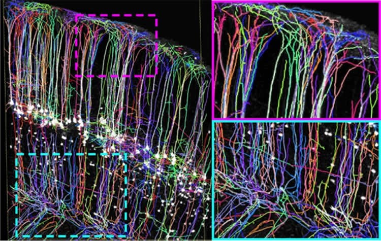
The uDISCO procedure had already permitted Ertürk and his group to obtain detailed views of the intact nervous systems of the mouse. However, clarification of the tissues was incomplete, such that traces of bones and other tissues were still visible. For vDISCO, the mixture of chemicals used to clear tissues has been modified, yielding the first technique that is capable of making whole animals fully transparent.
The new procedure thus allows one to visualize and characterize the full complexity of the nervous systems of small mammals, and will also allow researchers to trace the cellular interactions that lead to inflammation and wound healing, for example. According to the news article in Nature, vDisko has already revealed unexpected structural connections between remote organs – a discovery which, among other things, could lead to new treatments for stroke patients.
Source: LMU
Publisher: Organized by NeuroscienceNews.com.
Image Source: NeuroscienceNews.com image is credited to erturklab.
Original Research: Abstract for “Panoptic imaging of transparent mice reveals whole-body neuronal projections and skull–meninges connections” by Ruiyao Cai, Chenchen Pan, Alireza Ghasemigharagoz, Mihail Ivilinov Todorov, Benjamin Förstera, Shan Zhao, Harsharan S. Bhatia, Arnaldo Parra-Damas, Leander Mrowka, Delphine Theodorou, Markus Rempfler, Anna L. R. Xavier, Benjamin T. Kress, Corinne Benakis, Hanno Steinke, Sabine Liebscher, Ingo Bechmann, Arthur Liesz, Bjoern Menze, Martin Kerschensteiner, Maiken Nedergaard & Ali Ertürk in Nature Neuroscience. Published January 2 2019.
doi:10.1038/s41593-018-0301-3
[cbtabs][cbtab title=”MLA”]LMU”Imaging Nerve Cell Interactions.” NeuroscienceNews. NeuroscienceNews, 2 January 2019.
<https://neurosciencenews.com/neuron-interaction-imaging-10412/>.[/cbtab][cbtab title=”APA”]LMU(2019, January 2). Imaging Nerve Cell Interactions. NeuroscienceNews. Retrieved January 2, 2019 from https://neurosciencenews.com/neuron-interaction-imaging-10412/[/cbtab][cbtab title=”Chicago”]LMU”Imaging Nerve Cell Interactions.” https://neurosciencenews.com/neuron-interaction-imaging-10412/ (accessed January 2, 2019).[/cbtab][/cbtabs]
Abstract
Panoptic imaging of transparent mice reveals whole-body neuronal projections and skull–meninges connections
Analysis of entire transparent rodent bodies after clearing could provide holistic biological information in health and disease, but reliable imaging and quantification of fluorescent protein signals deep inside the tissues has remained a challenge. Here, we developed vDISCO, a pressure-driven, nanobody-based whole-body immunolabeling technology to enhance the signal of fluorescent proteins by up to two orders of magnitude. This allowed us to image and quantify subcellular details through bones, skin and highly autofluorescent tissues of intact transparent mice. For the first time, we visualized whole-body neuronal projections in adult mice. We assessed CNS trauma effects in the whole body and found degeneration of peripheral nerve terminals in the torso. Furthermore, vDISCO revealed short vascular connections between skull marrow and brain meninges, which were filled with immune cells upon stroke. Thus, our new approach enables unbiased comprehensive studies of the interactions between the nervous system and the rest of the body.


