Summary: Researchers have created a 3D atlas of the developing mouse brain, offering a dynamic, high-resolution view of brain structures during embryonic and post-natal stages. This new tool allows scientists to explore how brain cells, such as GABAergic neurons linked to neurological disorders, emerge and interact during development.
By integrating MRI and light sheet fluorescence microscopy, the atlas provides a reference framework for studying neurodevelopmental disorders and advancing neuroscience research. The atlas is available online, offering global access to this essential resource for brain research.
Key Facts:
- A 3D atlas maps brain development across seven stages in mice.
- The atlas tracks GABAergic neurons, key in disorders like autism and schizophrenia.
- It offers a free, interactive tool for researchers to explore neurodevelopment.
Source: Penn State
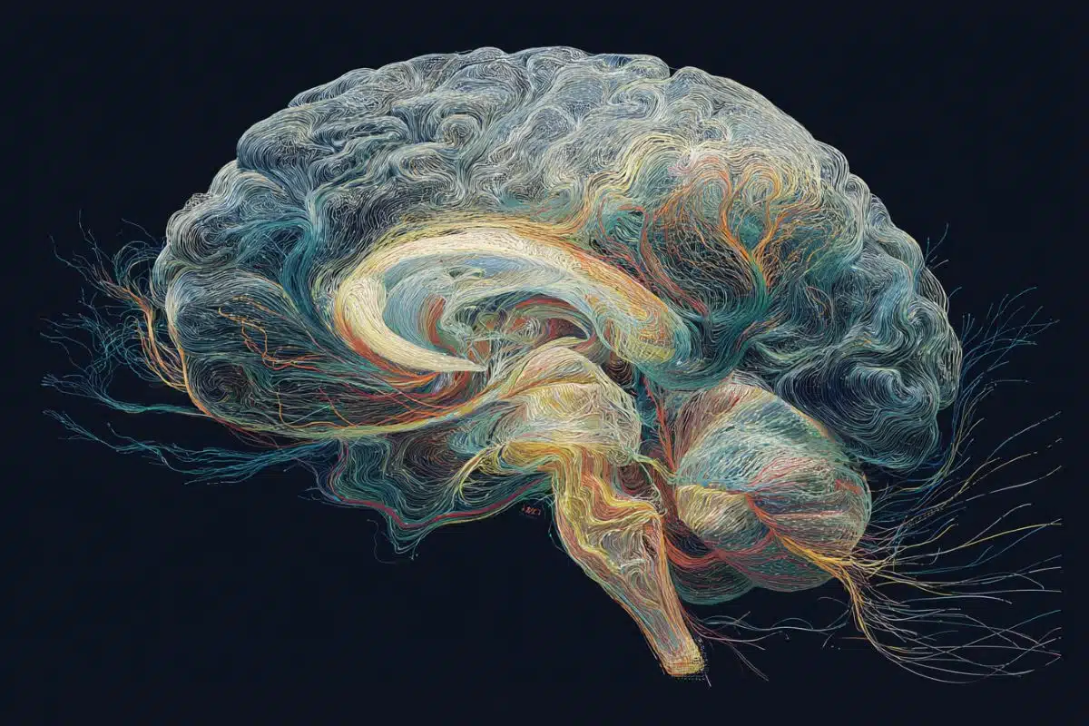
A 3D atlas of developing mice brains using advanced imaging and microscopy techniques has been created by a team of researchers at Penn State College of Medicine and collaborators from five different institutes.
This new atlas provides a more dynamic, 360-degree picture of the whole mammalian brain as it develops during the embryonic and immediate post-natal stages and serves as a common reference and anatomical framework that will help researchers understand brain development and study neurodevelopmental disorders.

They published their work today (Oct 21) in Nature Communications.
“Maps are a fundamental infrastructure to build knowledge upon but we don’t have a high-resolution 3D atlas of the developing brain,” said Yongsoo Kim, associate professor of neural and behavioral sciences at Penn State College of Medicine and senior author on the paper.
“We are generating high-resolution maps that we can use to understand how the brain grows under normal circumstances and what happens when a brain disorder emerges.”
Geographical atlases are a collection of maps that provide a comprehensive view of the Earth’s geography including boundaries between regions and countries, features like mountains and rivers, and thoroughfares like roads and highways. Importantly, they provide a common understanding that allows users pinpoint specific locations and understand the spatial relationship between regions.
Similarly, brain atlases are foundational for understanding the architecture of the brain. They help researchers visualize how the brain is organized spatially and understand brain structure, function, and how different regions and neurons are connected. Previously, scientists have been limited to 2D histology-based snapshots, which makes it challenging to interpret anatomical regions in three dimensions and any changes that may occur, Kim said.
In recent years, there has been tremendous progress in whole brain imaging techniques that let researchers look at the whole brain at high resolution and produce large-scale 3D datasets. To analyze this data, Kim explained, scientists have developed 3D reference atlases of the adult mouse brain, which is a model for the mammalian brain.
The atlases provide a universal anatomical framework that allow researchers to overlay diverse datasets and conduct comparative analyses. However, there’s no equivalent for the developing mouse brain, which undergoes rapid changes in shape and volume during the embryonic and post-natal stages.
“Without this 3D map of the developing brain, we cannot integrate data from emerging 3D studies into a standard spatial framework or analyze the data in a consistent manner,” Kim said. In other words, the lack of a 3D map hinders the advancement of neuroscience research.
The research team created a multimodal 3D common coordinate framework of the mouse brain across seven developmental timepoints — four points of time during the embryonic period and three periods during the immediate postnatal phase. Using MRI, they captured images of the brain’s overall form and structure.
They then employed light sheet fluorescence microscopy, an imaging technique that enables visualization of the whole brain at a single-cell resolution. These high-resolution images were then matched to the shape of the MRI templates of the brain to create the 3D map. The team pooled samples from both male and female mice.
To demonstrate how the atlas can be used to analyze different datasets and track how individual cell types emerge in the developing brain, the team focused on GABAergic neurons, which are nerve cells that play a key communication role in the brain. This cell type has been implicated in schizophrenia, autism and other neurological disorders.
While scientists have studied GABAergic neurons in the outermost region of the brain called the cortex, not much is known about how these cells arise in the whole brain during development, according to the researchers.
Understanding how these clusters of cells develop under normal conditions may be key to assessing what happens when something goes awry.
To facilitate collaboration and further advancement in neuroscience research, the team created an interactive web-based version that is publicly available and free. The aim is to significantly lower technical barriers for researchers around the world to access this resource.
“This provides a roadmap that can integrate a lot of different data — genomic, neuroimaging, microscopy and more — into the same data infrastructure. It will drive the next evolution of brain research driven by machine learning and artificial intelligence,” Kim said.
Other Penn State College of Medicine authors on the paper include: Fae Kronman, joint degree student in the MD/PhD Medical Scientist Training Program; Josephine Liwang, doctoral student; Rebecca Betty, research technologist; Daniel Vanselow, research project manager; Steffy Manjila, postdoctoral scholar; Jennifer Minteer, research technologist; Donghui Shin, research technologist; Rohan Patil, student; and Keith Cheng, distinguished professor, department of pathology.
Nicholas Tustison at the University of Virginia School of Medicine; Ashwin Bhandiwad and Lydia Ng at the Allen Institute for Brain Science; Choong Heon Lee and Jiangyang Zhang at the NYU Grossman School of Medicine; Jeffrey Duda and James Gee at the University of Pennsylvania; Jian Xue and Yingxi Lin at the University of Texas Southwestern Medical Center; Luis Puelles at the Universidad de Murcia; and Yuan-Ting Wu, who was previously research scientist at Penn State and currently project scientist at Cedars-Sinai Medical Center, also contributed to the paper.
Funding: The National Institutes of Health’s grants RF1MH12460501 from the Brain Research through Advancing Innovative Neurotechnologies (BRAIN) Initiative, R01NS108407, R01MH116176 and R01EB031722 supported this work.
About this brain development research news
Author: Christine Yu
Source: Penn State
Contact: Christine Yu – Penn State
Image: The image is credited to Neuroscience News
Original Research: Open access.
“Developmental mouse brain common coordinate framework” by Yongsoo Kim et al. Nature Communications
Abstract
Developmental mouse brain common coordinate framework
3D brain atlases are key resources to understand the brain’s spatial organization and promote interoperability across different studies. However, unlike the adult mouse brain, the lack of developing mouse brain 3D reference atlases hinders advancements in understanding brain development.
Here, we present a 3D developmental common coordinate framework (DevCCF) spanning embryonic day (E)11.5, E13.5, E15.5, E18.5, and postnatal day (P)4, P14, and P56, featuring undistorted morphologically averaged atlas templates created from magnetic resonance imaging and co-registered high-resolution light sheet fluorescence microscopy templates.
The DevCCF with 3D anatomical segmentations can be downloaded or explored via an interactive 3D web-visualizer. As a use case, we utilize the DevCCF to unveil GABAergic neuron emergence in embryonic brains. Moreover, we map the Allen CCFv3 and spatial transcriptome cell-type data to our stereotaxic P56 atlas.
In summary, the DevCCF is an openly accessible resource for multi-study data integration to advance our understanding of brain development.