Summary: For patients with retinitis pigmentosa, the gradual loss of vision can feel like an unstoppable clock. However, a new study has unlocked the biological “how” behind a promising cell-based therapy. By using single-cell analysis, researchers discovered that transplanted neural stem cells don’t just replace dead tissue; they act as a sophisticated repair crew.
These cells preserve vision by providing protective proteins, reducing cellular stress, and physically restoring the retina to a healthier state. In animal models, these transplants halted vision loss for a period equivalent to 20 human years, offering a new blueprint for treating degenerative eye diseases.
Key Facts
- Multifaceted Repair: Neural stem cells protect vision through four main mechanisms: delivering protective proteins, reducing stress in host cells, restoring retinal health, and maintaining structural integrity.
- Long-Term Protection: In rat models of retinitis pigmentosa, a single transplant preserved vision for up to 180 days (roughly 20 years in human terms).
- Dynamic Interaction: The study revealed that the relationship between transplanted stem cells and host retinal cells changes over time, adapting to the environment of the eye.
- Single-Cell Resolution: Researchers used advanced single-cell analysis to track exactly how the transplants “remodeled” the transcriptomic landscape of the diseased eye.
- Next Steps: Scientists are already engineering “super-stem cells” designed to express specific protective proteins identified in this study for even more powerful results.
Source: Cedars Sinai
Cedars-Sinai investigators working to optimize a cell-based treatment for retinitis pigmentosa have uncovered how transplanted neural stem cells interact with host retinal cells to preserve vision.
The findings, published in Nature Communications, may guide future research toward strategies to treat degenerative eye disease.

“We used single-cell analysis to show that neural stem cells can protect vision in several ways, including providing protective proteins, restoring retinal cells to a healthier state, reducing cellular stress, and maintaining retinal integrity,” said Clive Svendsen, PhD, executive director of the Board of Governors Regenerative Medicine Institute and co-corresponding author of the study.
Investigators transplanted neural stem cells into the retinas—the light-sensitive tissue lining the back of the eye—of laboratory rats with retinal degeneration. Previous studies have shown the transplants significantly reduced vision loss in the animals for up to 180 days, the equivalent of about 20 years in humans. In this study the team examined interactions between the transplanted cells and diseased retinal cells to better understand the neural stem cells’ protective effects.
“Our study reveals that the interaction between neural stem cells and host retinal cells dynamically changes over time,” said Shaomei Wang, MD, PhD, professor of Biomedical Sciences and co-corresponding author of the study. “Through a better understanding of this process, we may be able to develop more powerful approaches to treat eye diseases in the future.”
Investigators are now evaluating the use of neural stem cells engineered to express key protective proteins identified in this study to further improve the host retinal environment.
Additional Cedars-Sinai authors include Saba Shahin, Shaughn Bell, Bin Lu, Hui Xu, Jason Chetsawang, Stephany Ramirez, Jorge S. Alfaro, Alexander Laperle and Soshana Svendsen.
Other authors include Somanshu Banerjee and Vivek Swarup.
Funding: This work was supported by the California Institute Regenerative Medicine (LSP1-08235). J.C. was supported by CIRM-EDUC-08383 and S.R. was supported by CIRM-EDUC2-12638, and funding from the Board of Governors Regenerative Medicine Institute at Cedars-Sinai Medical Center.
Key Questions Answered:
A: While “cure” is a strong word, this research shows we can drastically slow or even halt the progression of blindness in degenerative diseases. Instead of just replacing old cells, these neural stem cells “nurse” the remaining host cells back to health, keeping the vision you have for much longer—potentially decades.
A: The study found that the stem cells are “dynamic.” They don’t just sit there; they continuously interact with the host retina, providing a steady supply of protective proteins and reducing the toxic stress that normally causes retinal cells to die. It’s like installing a permanent, self-adjusting maintenance crew inside the eye.
A: Clinical researchers are already looking at how to move this from animal models to human trials. By identifying the exact proteins that these cells use to protect the eye, they are now creating even more effective versions of the therapy that could be ready for testing in the near future.
Editorial Notes:
- This article was edited by a Neuroscience News editor.
- Journal paper reviewed in full.
- Additional context added by our staff.
About this genetics and visual neuroscience research news
Author: Cara Martinez
Source: Cedars Sinai
Contact: Cara Martinez – Cedars Sinai
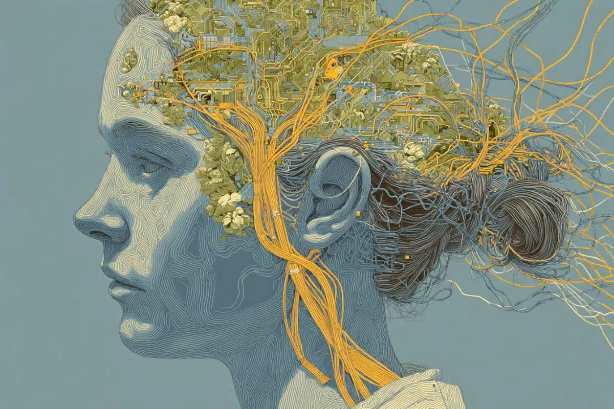
Image: The image is credited to Neuroscience News
Original Research: Open access.
“Dynamic transcriptomic remodeling in grafted human neural progenitor cells uncovers mechanisms for vision preservation in a rat model of retinitis pigmentosa” by Saba Shahin, Shaughn Bell, Bin Lu, Somanshu Banerjee, Vivek Swarup, Hui Xu, Jason Chetsawang, Stephany Ramirez, Jorge S. Alfaro, Alexander Laperle, Soshana Svendsen, Clive N. Svendsen & Shaomei Wang. Nature Communications
DOI:10.1038/s41467-026-69776-4
Abstract
Dynamic transcriptomic remodeling in grafted human neural progenitor cells uncovers mechanisms for vision preservation in a rat model of retinitis pigmentosa
Human neural progenitor cells (hNPCs) show promise in slowing retinal degeneration and are currently being tested in clinical trials for retinitis pigmentosa (RP). However, the long-term fate of grafted hNPCs and their interaction with host retinal cells remain unclear.
Here, we used single-cell transcriptomics to investigate temporal gene expression changes in grafted hNPCs and host retinal cells following subretinal injection into a rodent model of RP, revealing dynamic transcriptomic changes in the degenerative retinal environment.
Grafted hNPCs primarily differentiate into an astroglial phenotype and mature over time, contributing to photoreceptor protection through trophic factor secretion, metabolic modulation, suppression of apoptosis, oxidative stress, and inflammation, alongside extracellular matrix remodeling.
CellChat analysis revealed a decline in intercellular signaling and communication strength over time, correlating with weakened hNPC-host interactions.
These findings suggest that enhancing trophic factor support, particularly MANF, and improving the host retinal environment are critical for sustaining long-term vision preservation.