

Summary: Researchers observe the action and plot the movement of neurons with the help of a new technique.
Source: University of Queensland.
A University of Queensland team is among the first in neuroscience to see the brain’s tiniest molecules in action and plot their movements.
Professor Fred Meunier’s laboratory at the Clem Jones Centre for Ageing Dementia Research at the Queensland Brain Institute has developed a breakthrough technique in super-resolution microscopy.
“The technique will be revolutionary for neuroscientists and cell biologists,” Professor Meunier said.
“This is an exciting time, as we’ve opened the door for many more ground-breaking studies that will change our view of how molecules function to make the brain work.
“These images could further our understanding of memory, learning, and neurodegenerative diseases.”
The breakthrough imaging technique captures the movement of molecules within synapses – structures connecting brain cells – that are too small for conventional microscopes.
Classical optical microscopes can only clearly identify objects that are 200 nanometres (1/5000th of a millimetre).
Super-resolution microscopy techniques can achieve 10 to 20 times higher resolution (closer to electron microscopy), even on living cells.
Professor Meunier’s team has gone one step further by extending super-resolution capabilities to explore the dynamics of the nanoworld of our brain cells, including:
- Tracking single molecules in living brain cells undergoing communication
- Tracking these molecules within brain cells of living organisms such as fruit flies
- Tracking single tiny structures, called synaptic vesicles, that are responsible for the release of neurotransmitters at the synapse
- Imaging tiny signalling endosomes responsible for brain cell survival as they move from the synapse to the cell body
“We can now see molecules organise themselves by viewing the nitty gritty of mechanisms that allow neurons to communicate,” Professor Meunier said.
“You see molecules moving randomly and then, for a few milliseconds, interacting with each other.
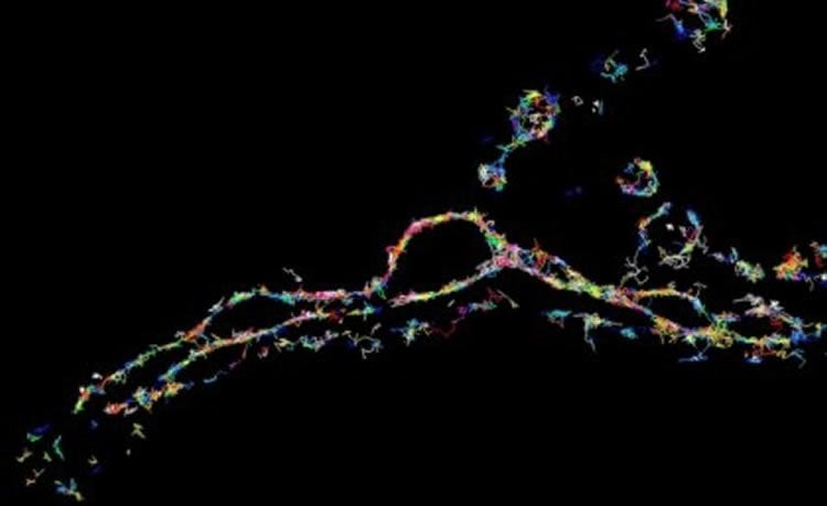
“Control of these critical moments define neurotransmission and allow brain cells to communicate efficiently.”
The technique has led to a series of papers from Professor Meunier’s team published in the Journal of Cell Biology and Nature Communications.
Funding: The research was funded by the Australian Research Council and the National Health and Medical Research Council.
Source: University of Queensland
Image Source: NeuroscienceNews.com image is adapted from the University of Queensland press release.
Original Research: The studies appear in Journal of Cell Biology and Nature Communications.
[cbtabs][cbtab title=”MLA”]University of Queensland “Sneak Peek Into the Nano World of Brain Cells.” NeuroscienceNews. NeuroscienceNews, 9 January 2017.
<https://neurosciencenews.com/nanosciene-neuroscience-neurons-5896/>.[/cbtab][cbtab title=”APA”]University of Queensland (2017, January 9). Sneak Peek Into the Nano World of Brain Cells. NeuroscienceNew. Retrieved January 9, 2017 from https://neurosciencenews.com/nanosciene-neuroscience-neurons-5896/[/cbtab][cbtab title=”Chicago”]University of Queensland “Sneak Peek Into the Nano World of Brain Cells.” https://neurosciencenews.com/nanosciene-neuroscience-neurons-5896/ (accessed January 9, 2017).[/cbtab][/cbtabs]


