Summary: Researchers have implicated mossy cells in both seizures and memory problems in those with temporal lobe epilepsy.
Source: NIH/NINDS.
A small group of cells in the brain can have a big effect on seizures and memory in a mouse model of epilepsy. According to a new study in Science, loss of mossy cells may contribute to convulsive seizures in temporal lobe epilepsy (TLE) as well as memory problems often experienced by people with the disease. The study was funded by the National Institute of Neurological Disorders and Stroke (NINDS), part of the National Institutes of Health.
“The role of mossy cells in epilepsy has been debated for decades. This study reveals how critical these cells are in the disease, and the findings suggest that preventing loss of mossy cells or finding ways to activate them may be potential therapeutic targets,” said Vicky Whittemore, Ph.D., program director at NINDS.
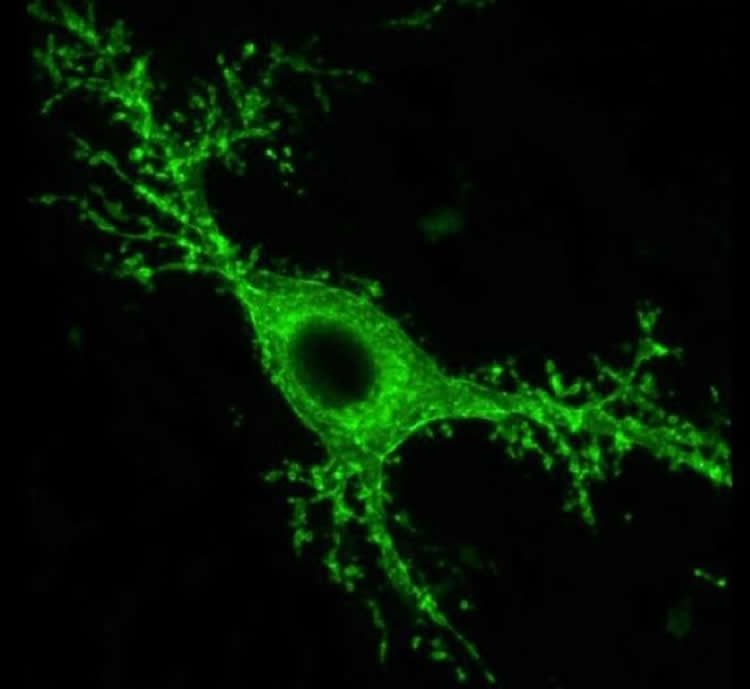
Mossy cells, named for the dense moss-like protrusions that cover their surface, are located in the hippocampus, a brain area that is known to play key roles in memory. Loss of mossy cells is associated with TLE, but it is unknown what role that plays in the disease. Using state-of-the-art tools, Ivan Soltesz, Ph.D., professor of neurosurgery and neurosciences at Stanford University, Palo Alto, California, and his team were able to turn mossy cells on and off to track their effects in a mouse model of epilepsy.
“This study would not have been possible without the rapid advancement of technology, thanks in part to the BRAIN Initiative, which has encouraged scientists to develop innovative instruments and new ways to look at the brain,” said Dr. Soltesz. “It’s remarkable that we can manipulate specific brain cells in the hippocampus of a mouse. Using 21st century tools brings us closer than ever to unlocking the mysteries behind this debilitating disease.”
In TLE, many seizures, known as focal seizures, originate in one part of the brain and are evident on electroencephalography (EEG) scans that show the brain’s electrical activity. These seizures can result in symptoms such as twitching or a strange taste or smell, and many people with TLE might not be aware that these symptoms are seizures. Sometimes, focal seizures can spread throughout the entire brain becoming generalized, resulting in involuntary muscle spasms, or convulsions, that affect the limbs and other parts of the body as well as loss of consciousness.
When Dr. Soltesz’ group detected focal seizures on the mice’s EEG scans, they turned mossy cells on or off to see whether they had any effect on the seizures. The researchers found that turning on the cells prevented the focal seizures from transitioning into convulsive ones. When the mossy cells were turned off, however, convulsive seizures were more likely to occur. Mossy cells had only a minor effect on the occurrence of focal seizures.
“This was the first time we were able to show specifically that mossy cell activity can control convulsive seizures,” said Anh Bui, an M.D., Ph.D. student at the University of California-Irvine, and first author of the paper. “These mice were missing most of their mossy cells, yet we were able to see effects just by manipulating the small number of surviving cells.”
People with TLE often experience temporary changes in thinking and long-term problems with memory. Dr. Soltesz and his colleagues looked at the role of mossy cells in two specific types of memory: object recognition and spatial memory, which refers to identifying where objects are located and navigating around the environment. In these experiments, the mice were placed in a chamber with two identical items. The following day, one of the items was replaced with a different one (to test for object recognition) or moved to a different location (to test for spatial memory).
The epileptic mice had trouble with spatial memory tasks but their ability to recognize objects was unaffected. In addition, turning off mossy cells in healthy mice also led to problems with spatial memory in those animals. These findings suggest that a decrease in mossy cells may lead to convulsive seizures as well as memory deficits.
More research is needed to further understand the role of mossy cells in seizure progression as well as their effects early in the disease.
Funding: This work was supported by the NINDS (NS086429, NS074702, NS094668).
Source: Barbara McMakin – NIH/NINDS
Publisher: Organized by NeuroscienceNews.com.
Image Source: NeuroscienceNews.com image is credited to Ivan Soltesz, Ph.D., Stanford University.
Original Research: Abstract in Science.
doi:10.1126/science.aan4074
[cbtabs][cbtab title=”MLA”]NIH/NINDS “Epilepsy Study Links Mossy Brain Cells to Seizures and Memory Loss.” NeuroscienceNews. NeuroscienceNews, 16 February 2018.
<https://neurosciencenews.com/mossy-cells-epilepsy-memory-8507/>.[/cbtab][cbtab title=”APA”]NIH/NINDS (2018, February 16). Epilepsy Study Links Mossy Brain Cells to Seizures and Memory Loss. NeuroscienceNews. Retrieved February 16, 2018 from https://neurosciencenews.com/mossy-cells-epilepsy-memory-8507/[/cbtab][cbtab title=”Chicago”]NIH/NINDS “Epilepsy Study Links Mossy Brain Cells to Seizures and Memory Loss.” https://neurosciencenews.com/mossy-cells-epilepsy-memory-8507/ (accessed February 16, 2018).[/cbtab][/cbtabs]
Abstract
Dentate gyrus mossy cells control spontaneous convulsive seizures and spatial memory
Temporal lobe epilepsy (TLE) is characterized by debilitating, recurring seizures and an increased risk for cognitive deficits. Mossy cells (MCs) are key neurons in the hippocampal excitatory circuit, and the partial loss of MCs is a major hallmark of TLE. We investigated how MCs contribute to spontaneous ictal activity and to spatial contextual memory in a mouse model of TLE with hippocampal sclerosis, using a combination of optogenetic, electrophysiological, and behavioral approaches. In chronically epileptic mice, real-time optogenetic modulation of MCs during spontaneous hippocampal seizures controlled the progression of activity from an electrographic to convulsive seizure. Decreased MC activity is sufficient to impede encoding of spatial context, recapitulating observed cognitive deficits in chronically epileptic mice.