

Summary: Researchers have singled and taken control of a circuit in mouse brains in order to dial the animal’s mood up or down.
Source: Duke University.
By combining super-fine electrodes and tiny amounts of a very specific drug, Duke University researchers have singled out a circuit in mouse brains and taken control of it to dial an animal’s mood up and down.
Stress-susceptible animals that behaved as if they were depressed or anxious were restored to relatively normal behavior by tweaking the system, according to a study appearing in the July 20 issue of Neuron.
“If you ‘turn the volume up’ on animals that hadn’t experienced stress, they start normal and then they have a problem,” said lead researcher Kafui Dzirasa, an assistant professor of psychiatry and behavioral sciences, and neurobiology. “But in the animals that had experienced stress and didn’t do well with it, you had to turn their volume up to get them back to normal. It looked like stress had turned the volume down.”
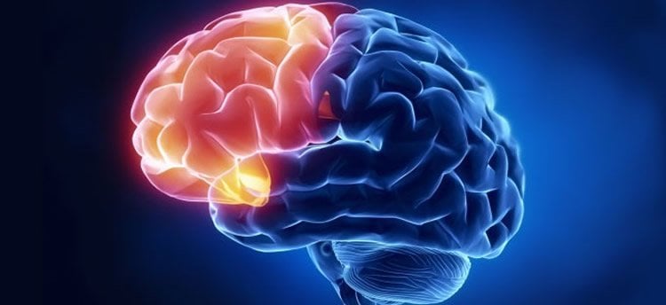
The circuit the team identified and altered is a connection the prefrontal cortex uses to keep time for the limbic system, which governs emotions and basic drives. To regulate mood, the prefrontal cortex acts as a pacemaker to coordinate the actions of the amygdala, which governs stress responses, and the ventral tegmental area, which plays a role in the brain’s reward circuitry.
“These subcortical circuits are the key regulators of our emotional life,” said Helen Mayberg, a professor of psychiatry, neurology and radiology at Emory University who was not involved in this research. “What’s great about this paper is that they use different approaches to see a circuit that’s relevant to a lot of disorders,” said Mayberg, who has been pioneering deep-brain stimulation of very specific sites in the human prefrontal cortex to treat mood disorders.
The emerging picture from this study and others is of a brain built of multi-part circuits that respond in concert and regulate one another. Specificity in understanding these circuits is going to be key to resolving different disorders, Dzirasa said.
“The prefrontal cortex is not just a blob of cells,” Mayberg said. “These findings give insight into which cells go to which area and allow researchers to kind of choreograph their actions.”
Dzirasa is an M.D. just finishing his residency in psychiatry and a Ph.D. neuroscientist with an engineering background. Postdoctoral researcher and first author Rainbo Hultman is a biochemist.
In addition to overcoming the challenges of understanding each other, they asked, “Could we go from a protein, to a signaling activity, to a cell, to a circuit, to this big activity that happens across the whole brain, to actual behavior?” Hultman said.
“Illness can happen at any one of these levels,” said Dzirasa, who is also a member of the Duke Institute for Brain Sciences.
The team started by precisely placing arrays of 32 electrodes in four brain areas of the mice. Then they recorded brain activity as these mice were subjected to a stressful situation called chronic social defeat. This allowed them to see activity between the prefrontal cortex and three areas of the limbic system that are implicated in major depression.
To interpret the complicated data coming from the electrodes, the neuroscientists then turned to Duke colleagues David Dunson of statistical science and Lawrence Carin of electrical engineering, who specialize in statistical analysis of noisy data to find important patterns. Using machine learning algorithms, they identified which parts of the data seemed to be the timing control signal between the prefrontal cortex and the amygdala and zeroed in on the individual neurons involved in that circuit.
“They came back with, ‘It’s this clock signature here that is responsible for which mice become susceptible to stress and which become resilient,'” Dzirasa said.
Hultman then turned to engineered molecules called DREADD developed by University of North Carolina at Chapel Hill pharmacologist Bryan Roth. These Designer Receptors Exclusively Activated by Designer Drug are very specific signal receptors that can be incorporated into the neural circuit’s control spots in very tiny amounts (0.5 microliter). A drug that attaches only to that DREADD is then administered to give the researchers control over the circuit.
This new combination of electronics and drugs to intervene in an individual brain circuit might be used to create mouse models of other mood disorders for other studies, Dzirasa said. But Emory’s Mayberg cautions that a mouse brain is not a human brain and to assess anything like “mood” in a mouse, one can only infer from its behaviors. “It’s hard to do, even in a human,” she said.
Funding: This work was supported by funding from National Institutes of Mental Health grants R37MH073853 and R01MH099192, and a research incubator award from the Duke Institute for Brain Sciences.
Source: Karl Leif Bates – Duke University
Image Source: This NeuroscienceNews.com image is in the public domain.
Original Research: Abstract for “Dysregulation of Prefrontal Cortex-Mediated Slow-Evolving Limbic Dynamics Drives Stress-Induced Emotional Pathology” by Rainbo Hultman, Stephen D. Mague, Qiang Li, Brittany M. Katz, Nadine Michel, Lizhen Lin, Joyce Wang, Lisa K. David, Cameron Blount, Rithi Chandy, David Carlson, Kyle Ulrich, Lawrence Carin, David Dunson, Sunil Kumar, Karl Deisseroth, Scott D. Moore, and Kafui Dzirasa in Neuron. Published online June 23 2016 doi:10.1016/j.neuron.2016.05.038
[cbtabs][cbtab title=”MLA”]Duke University. “Precise Control of Brain Circuits Alters Mood: Mouse Study.” NeuroscienceNews. NeuroscienceNews, 23 June 2016.
<https://neurosciencenews.com/mood-neural-networks-4553/>.[/cbtab][cbtab title=”APA”]Duke University. (2016, June 23). Precise Control of Brain Circuits Alters Mood: Mouse Study. NeuroscienceNews. Retrieved June 23, 2016 from https://neurosciencenews.com/mood-neural-networks-4553/[/cbtab][cbtab title=”Chicago”]Duke University. “Precise Control of Brain Circuits Alters Mood: Mouse Study.” https://neurosciencenews.com/mood-neural-networks-4553/ (accessed June 23, 2016).[/cbtab][/cbtabs]
Abstract
Dysregulation of Prefrontal Cortex-Mediated Slow-Evolving Limbic Dynamics Drives Stress-Induced Emotional Pathology
Highlights
•Prefrontal cortex (PFC) oscillations synchronize with ultraslow limbic dynamics
•PFC unit firing signals the synchronization state of amygdala (AMY) and ventral tegmental area (VTA)
•Chronic stress selectively disrupts PFC-dependent regulation of AMY-VTA synchrony
•PFC to AMY circuit stimulation recovers normal network function and behavior
Summary
Circuits distributed across cortico-limbic brain regions compose the networks that mediate emotional behavior. The prefrontal cortex (PFC) regulates ultraslow (<1 Hz) dynamics across these networks, and PFC dysfunction is implicated in stress-related illnesses including major depressive disorder (MDD). To uncover the mechanism whereby stress-induced changes in PFC circuitry alter emotional networks to yield pathology, we used a multi-disciplinary approach including in vivo recordings in mice and chronic social defeat stress. Our network model, inferred using machine learning, linked stress-induced behavioral pathology to the capacity of PFC to synchronize amygdala and VTA activity. Direct stimulation of PFC-amygdala circuitry with DREADDs normalized PFC-dependent limbic synchrony in stress-susceptible animals and restored normal behavior. In addition to providing insights into MDD mechanisms, our findings demonstrate an interdisciplinary approach that can be used to identify the large-scale network changes that underlie complex emotional pathologies and the specific network nodes that can be used to develop targeted interventions.
“Dysregulation of Prefrontal Cortex-Mediated Slow-Evolving Limbic Dynamics Drives Stress-Induced Emotional Pathology” by Rainbo Hultman, Stephen D. Mague, Qiang Li, Brittany M. Katz, Nadine Michel, Lizhen Lin, Joyce Wang, Lisa K. David, Cameron Blount, Rithi Chandy, David Carlson, Kyle Ulrich, Lawrence Carin, David Dunson, Sunil Kumar, Karl Deisseroth, Scott D. Moore, and Kafui Dzirasa in Neuron. Published online June 23 2016 doi:10.1016/j.neuron.2016.05.038


