

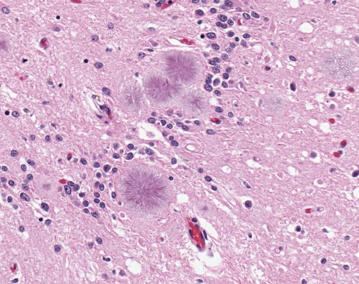
The plaque deposits in the brain of Alzheimer’s patients are surrounded by the brain’s own immune cells, the microglia. This was already recognized by Alois Alzheimer more than one hundred years ago. But until today it still remains unclear what role microglia play in Alzheimer’s disease. Do they help to break down the plaque deposit?
A study by researchers of the Max Delbrück Center for Molecular Medicine (MDC) Berlin-Buch and Charité – Universitätsmedizin Berlin has now shed light on these mysterious microglia during the progression of Alzheimer’s disease.
Dr. Grietje Krabbe of the laboratory of Professor Helmut Kettenmann (MDC) and Dr. Annett Halle of the Neuropathology Department of the Charité headed by Professor Frank Heppner demonstrated that the microglial cells around the deposits do not show the classical activation pattern in mouse models of Alzheimer´s disease. On the contrary, in the course of the Alzheimer’s disease they lose two of their biological functions. Both their ability to remove cell fragments or harmful structures and their directed process motility towards acute lesions are impaired. The impact of the latter loss-of-function needs further investigation. The plaques consist of protein fragments, the beta-amyloid peptides, which in Alzheimer’s disease are deposited in the brain over the course of years. They are believed to be involved in destroying the nerve cells of the affected patients, resulting in an incurable cognitive decline.

However, just why the microglial cells, which cluster around the deposits, are inactivated or lose their functionality is still not fully understood. The researchers concluded that this process occurs at a very early stage of disease development and is likely triggered by the beta-amyloid. This is confirmed by the fact that the loss-of-function of the microglial cells in the mice could be reversed by beta-amyloid antibodies thereby decreasing the beta-amyloid burden. According to the researchers, the potential to restore microglial function by directed manipulation should be pursued and exploited to develop treatments for Alzheimer’s disease.
Notes about this Alzheimer’s disease research
Contact: Barbara Bachtler – Max Delbrück Center for Molecular Medicine (MDC) Berlin-Buch in the Helmholtz Association
Source: Max Delbrück Center for Molecular Medicine press release
Image Source: The beta-amyloid plaque image is credited to the CDC and is available in the public domain.
Original Research: Full open access research for “Functional Impairment of Microglia Coincides with Beta-Amyloid Deposition in Mice with Alzheimer-Like Pathology” by Grietje Krabber, Annett Halle, Vitali Matyash, Jan L. Rinnenthal, Gina D. Eom, Ulrike Bernhardt, Kelly R. Miller, Stefan Prokop, Helmut Kettenmann and Frank L. Heppner in PLoS ONE. Published online April 8 2013 doi:10.1371/journal.pone.0060921


