Summary: Researchers have identified a mechanism that keeps the brain clean during neurodegenerative diseases.
Source: PLOS.
Research led by the Achucarro Basque Center for Neuroscience, the University of the Basque Country (UPV/EHU), and the Ikerbasque Foundation has revealed the mechanisms that keep the brain clean during neurodegenerative diseases.
When neurons die, their debris need to be quickly removed in order for the surrounding brain tissue to continue to function properly. Elimination of the neuron corpses, in a process called phagocytosis, is carried out by highly specialized cells in the brain called microglia. These small cells have many ramifications that are in constant motion and are specially equipped to detect and destroy any foreign element, including dead neurons. Or so it was thought until now.
This study, publishing May 26, 2016 in PLOS Biology, investigates, for the first time, the process of neuronal death and microglial phagocytosis in the diseased brain. To this end, scientists collected brain samples from epilepsy patients at University Hospital of Cruces and from epileptic mice.
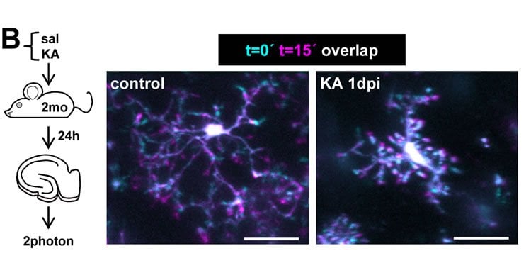
It is known that during epilepsy-associated seizures, neurons die. However, contrary to what happens in the healthy brain, during epilepsy, microglia seem to be “blind” and unable to find the dead neurons and to destroy them. Their behavior is abnormal. Therefore, dead neurons cannot be eliminated and accumulate, spreading the damage to neighboring neurons and triggering an inflammatory response that worsens the brain injury.
This discovery opens a new avenue to explore therapies that could alleviate the effects of brain diseases. In fact, the research group that undertook these studies is currently developing drugs, hoping to boost this cleaning process -phagocytosis- and help in the treatment of epilepsy.
The study was led by Dr. Amanda Sierra, director of the Laboratory of Glial Cell Biology at the Achucarro Basque Center for Neuroscience. The experimental work was mainly carried out by Oihane Abiega, Sol Beccari, and Irune Diaz Aparicio. Other scientists from Achucarro and UPV/EHU, including Juan Manuel Encinas, Jorge Valero, Victor Sanchez-Zafra, and Inaki Paris, also contributed to the study
This international research effort was coordinated from the Basque Country, and scientists from CIC bioGUNE (Spain), the University of Bordeaux (France), the University of Southampton (UK), Laval University (Canada), and Baylor College of Medicine (USA) also took part.
Funding: This work was supported by grants from the Spanish Ministry of Economy and Competitiveness with FEDER funds to AS (BFU2012-32089 and RYC-2013-12817) and JME (SAF2012-40085 and RYC-2012-11137); and from the Basque Government (Saiotek S-PC 12UN014) and Ikerbasque start-up funds to AS and JME; the Natural Sciences and Engineering Research Council of Canada NSERC (RGPIN-2014-05308) to MET; NIH Intellectual and Developmental Disabilities Research Grant (P30HD024064) and Dana Foundation and McKnight Endowment for Science Work grants to MMS; grants from NIH R01 NS, 39943 and 49427 to AEA; and T32 NS and 43124 to ALB, who is a recipient of an Epilepsy Foundation Postdoctoral Fellowship; and Medical Research Councilk; MR/K022687/1) to DGN. In addition, OA is recipient of a predoctoral fellowship from the Basque Government, IDA is recipient of a predoctoral fellowship from the University of the Basque Country EHU/UPV, and VSZ is recipient of a predoctoral fellowship from the Spanish Ministry of Economy and Competitiveness. The funders had no role in study design, data collection and analysis, decision to publish, or preparation of the manuscript.
Competing Interests: The authors have declared that no competing interests exist.
Source: Jamie Sagarduy – PLOS
Image Source: This NeuroscienceNews.com image is credited to Abiega et al./PLOS Biology.
Original Research: Full open access research for “Neuronal Hyperactivity Disturbs ATP Microgradients, Impairs Microglial Motility, and Reduces Phagocytic Receptor Expression Triggering Apoptosis/Microglial Phagocytosis Uncoupling” by Oihane Abiega, Sol Beccari, Irune Diaz-Aparicio, Agnes Nadjar, Sophie Layé, Quentin Leyrolle, Diego Gómez-Nicola, María Domercq, Alberto Pérez-Samartín, Víctor Sánchez-Zafra, Iñaki Paris, Jorge Valero, Julie C. Savage, Chin-Wai Hui, Marie-Ève Tremblay, Juan J. P. Deudero, Amy L. Brewster, Anne E. Anderson, Laura Zaldumbide, Lara Galbarriatu, Ainhoa Marinas, Maria dM. Vivanco, Carlos Matute, Mirjana Maletic-Savatic, Juan M. Encinas, and Amanda Sierra in PLOS Biology. Published online May 26 2016 doi:10.1371/journal.pbio.1002466
[cbtabs][cbtab title=”MLA”]PLOS. “The Brain Needs Cleaning to Stay Healthy.” NeuroscienceNews. NeuroscienceNews, 27 May 2016.
<https://neurosciencenews.com/microglia-phagocytosis-neuroscience-4328/>.[/cbtab][cbtab title=”APA”]PLOS. (2016, May 27). The Brain Needs Cleaning to Stay Healthy. NeuroscienceNews. Retrieved May 27, 2016 from https://neurosciencenews.com/microglia-phagocytosis-neuroscience-4328/[/cbtab][cbtab title=”Chicago”]PLOS. “The Brain Needs Cleaning to Stay Healthy.” https://neurosciencenews.com/microglia-phagocytosis-neuroscience-4328/ (accessed May 27, 2016).[/cbtab][/cbtabs]
Abstract
Neuronal Hyperactivity Disturbs ATP Microgradients, Impairs Microglial Motility, and Reduces Phagocytic Receptor Expression Triggering Apoptosis/Microglial Phagocytosis Uncoupling
Phagocytosis is essential to maintain tissue homeostasis in a large number of inflammatory and autoimmune diseases, but its role in the diseased brain is poorly explored. Recent findings suggest that in the adult hippocampal neurogenic niche, where the excess of newborn cells undergo apoptosis in physiological conditions, phagocytosis is efficiently executed by surveillant, ramified microglia. To test whether microglia are efficient phagocytes in the diseased brain as well, we confronted them with a series of apoptotic challenges and discovered a generalized response. When challenged with excitotoxicity in vitro (via the glutamate agonist NMDA) or inflammation in vivo (via systemic administration of bacterial lipopolysaccharides or by omega 3 fatty acid deficient diets), microglia resorted to different strategies to boost their phagocytic efficiency and compensate for the increased number of apoptotic cells, thus maintaining phagocytosis and apoptosis tightly coupled. Unexpectedly, this coupling was chronically lost in a mouse model of mesial temporal lobe epilepsy (MTLE) as well as in hippocampal tissue resected from individuals with MTLE, a major neurological disorder characterized by seizures, excitotoxicity, and inflammation. Importantly, the loss of phagocytosis/apoptosis coupling correlated with the expression of microglial proinflammatory, epileptogenic cytokines, suggesting its contribution to the pathophysiology of epilepsy. The phagocytic blockade resulted from reduced microglial surveillance and apoptotic cell recognition receptor expression and was not directly mediated by signaling through microglial glutamate receptors. Instead, it was related to the disruption of local ATP microgradients caused by the hyperactivity of the hippocampal network, at least in the acute phase of epilepsy. Finally, the uncoupling led to an accumulation of apoptotic newborn cells in the neurogenic niche that was due not to decreased survival but to delayed cell clearance after seizures. These results demonstrate that the efficiency of microglial phagocytosis critically affects the dynamics of apoptosis and urge to routinely assess the microglial phagocytic efficiency in neurodegenerative disorders.
“Neuronal Hyperactivity Disturbs ATP Microgradients, Impairs Microglial Motility, and Reduces Phagocytic Receptor Expression Triggering Apoptosis/Microglial Phagocytosis Uncoupling” by Oihane Abiega, Sol Beccari, Irune Diaz-Aparicio, Agnes Nadjar, Sophie Layé, Quentin Leyrolle, Diego Gómez-Nicola, María Domercq, Alberto Pérez-Samartín, Víctor Sánchez-Zafra, Iñaki Paris, Jorge Valero, Julie C. Savage, Chin-Wai Hui, Marie-Ève Tremblay, Juan J. P. Deudero, Amy L. Brewster, Anne E. Anderson, Laura Zaldumbide, Lara Galbarriatu, Ainhoa Marinas, Maria dM. Vivanco, Carlos Matute, Mirjana Maletic-Savatic, Juan M. Encinas, and Amanda Sierra in PLOS Biology. Published online May 26 2016 doi:10.1371/journal.pbio.1002466