

Summary:
Scientists have created the first comprehensive 3D “Lipid Brain Atlas” of the mouse brain, identifying 539 distinct “lipizones,” or chemical territories, defined solely by their lipid signatures. The high-resolution map reveals that lipids function like molecular postal codes, linking distant but structurally related regions, while also uncovering unexpected patchwork heterogeneity within white matter that gene-based mapping has historically missed.
Key Facts:
- 539 “Lipizones” Identified: By combining mass spectrometry imaging with new machine-learning algorithms to process 7 million data points across the whole brain, the team defined 539 distinct metabolic territories based solely on their unique combination of lipids.
- Molecular Postal Codes: The atlas shows that lipids are not randomly distributed; instead, they follow precise anatomical borders and, critically, can link a nerve cell body to the distant target regions reached by its fibers, providing a shared chemical address.
- Unexpected White Matter Heterogeneity: The findings demonstrate that seemingly uniform white matter is actually a patchwork of distinct chemical zones, revealing that myelin-making cells possess a biochemical diversity that was previously invisible to gene-based maps.
Source: EPFL (École Polytechnique Fédérale de Lausanne)
The human brain is frequently described in terms of its cellular architecture and electrical wiring, the complex network of neurons and synapses. However, much of the brain’s physical structure is actually made of lipids. These fatty molecules form the essential membranes of every neuron, wrap nerve fibers with insulating myelin, and play critical roles in how brain cells send and receive signals.
Despite their fundamental importance to biological function, lipids have long been a relative blind spot in neuroscience. While standard imaging and sequencing methods have allowed scientists to map genes and proteins in exquisite detail, distinguishing between different lipid molecules at high resolution across whole tissues has proven extraordinarily challenging.
“So scientists have had only a blurry picture of how lipids differ from one part of the brain to another,” says Giovanni D’Angelo, professor at EPFL and Kristian Gerhard Jebsen Foundation Chair in Nutrition and Metabolism. “That gap matters, because changes in brain lipids are increasingly linked to conditions from depression to Alzheimer’s disease.”
A Distinctive Chemical Map
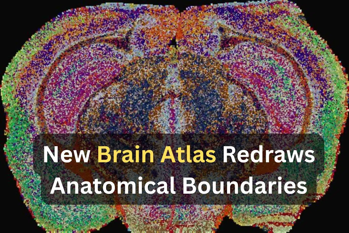
Now, an interdisciplinary team led by D’Angelo, Luca Fusar Bassini, and Gioele La Manno at EPFL has bridged this gap by constructing the first detailed, high-resolution atlas of lipids across the entire mouse brain. Published in Nature, the Lipid Brain Atlas demonstrates that these fatty molecules are distributed in a precise, orderly pattern that tightly lines up with the brain’s known anatomy.
Crucially, the lipid map revealed anatomical boundaries that structural imaging alone has historically missed. The signature was so distinctive that the researchers found they could identify the specific region of brain tissue simply by analyzing its lipid composition.
Building the Atlas: Mass Spectrometry Meets Machine Learning
To construct the atlas, the investigators utilized mass spectrometry imaging, a method that fires a laser at thin brain slices point-by-point to record exactly which lipids are situated at every spot. The team analyzed a total of 172 lipids across 109 individual slices taken from 11 mice.
Synthesizing roughly seven million individual readings into one coherent 3D model of the whole brain required the development of entirely new machine-learning tools, specifically engineered for the scale of this project.
The finished map divided the brain into 539 distinct territories, which the team labeled “lipizones,” each defined by its own signature biochemical mix.
While many lipizones correspond to established cell types and known brain regions, others provide entirely new relational links. Lipizones were found to connect clusters of nerve cell bodies directly to the far-off terminal areas that their fibers reach. This structural connection is one that gene-based maps, which typically focus on the cell bodies themselves, usually miss.
“In effect, lipids work like postal codes, giving distant but related parts of the brain the same chemical address,” D’Angelo explains.
White Matter’s Patchwork Chemistry and Pregnancy Retuning
The atlas also identified crucial patterns that earlier methods had entirely obscured. The brain’s white matter, historically thought of as a fairly uniform insulating material, emerged as a rich patchwork of distinct biochemical zones.
“This shows that the cells which make myelin are far more varied in their chemistry than their genes alone would suggest,” says Luca Fusar Bassini.
Furthermore, the team deployed the atlas to capture how the brain’s lipid landscape changes during pregnancy. In pregnant mice, lipids shifted dramatically across many regions, with changes larger overall than the baseline differences usually observed between males and females. The outer cortex was heavily reshaped, while a key building block of myelin called galactosyl ceramide rose sharply across much of the white matter.
“Together, these shifts suggest the brain retunes its own wiring to meet the demands of pregnancy,” says Gioele La Manno. “The Atlas reveals that the brain constantly adjusts its lipids to meet the body’s changing needs.”
Public Resource for Disease Research
The Lipid Brain Atlas is now freely available online as a public resource for the global research community. By providing a healthy reference map, it allows researchers to pinpoint where lipid metabolism goes wrong in neurodevelopmental, aging, and neurodegenerative disorders.
“What transfers to humans isn’t the map, it’s the method, and the fact that we now know that a healthy brain has a very organized lipid map at all,” Luca Fusar Bassini concludes.
Editorial Notes:
- This article was edited by a Neuroscience News editor.
- Journal paper reviewed in full.
- Additional context added by our staff.
About this Genetics and Neuroregeneration Research:
- Media Contact: Gioele La Manno
- Source: EPFL
- Image Credit: Image credited to EPFL/Luca Fusar Bassini
- Original Research is Open Access: Nature (September 23, 2026). “The lipidomic architecture of the mouse brain.” Authors: Luca Fusar Bassini, Halima Hannah Schede, Laura Capolupo, Leila Haj Abdullah Alieh, Irmak Kaysudu, Francesca Venturi, Hannah Hochgerner, Alessandro Valente, Colas Droin, Daniel Trejo Banos, Irina Khven, Jean Andrea Maillat, Anne-Laure Mahul-Mellier, Antonino Asaro, Doğukan H. Ülgen, Pavel Barahtjan, Ece Z. Asirim, Anita Nasrallah, Carmen Sandi, Ekaterina Krymova, Giovanni D’Angelo & Gioele La Manno.
- DOI: 10.1038/s41586-026-11050-0
Abstract
The lipidomic architecture of the mouse brain
Lipids are fundamental components of the brain, crucial for synaptic transmission and signal propagation. Altered brain lipid composition is associated with common and rare neuropathologies, yet the spatial organization of the mammalian brain lipidome remains insufficiently characterized compared with other modalities.
Here we mapped the membrane-lipid architecture of the adult mouse brain at micrometric scale, across sexes and during pregnancy. This lipid brain atlas reveals that lipids describe a fine-grained biochemical structure that aligns with functional anatomy.
Membrane-lipid spatial heterogeneity clusters into territories, which we termed ‘lipizones’. Lipizones partially mirror cell-type territories, but also capture distal axon terminals. Through lipizones, we (1) reveal the organizing principles of the grey matter lipidome, related to connectivity and cytoarchitecture; (2) discover a new axis of oligodendrocyte heterogeneity in the white matter; and (3) find biochemical zonation in the choroid plexus and in the ventricular walls.
We show that this lipidomic architecture can adapt to changing physiological needs. In the brain of pregnant female mice, the white matter is metabolically activated and the cortex undergoes a lipizone-specific remodelling that is particularly pronounced in layer 4.
These results are a foundational resource (https://lbae-v2.epfl.ch/) poised to reshape the understanding of lipids in brain development, physiology and pathology.


