Summary: Researchers have successfully recorded live images of leg regeneration of a crustacean following amputation.
Source: eLife.
Researchers have for the first time recorded how cells of the epidermis behave during the regrowth of adult limbs after amputation.
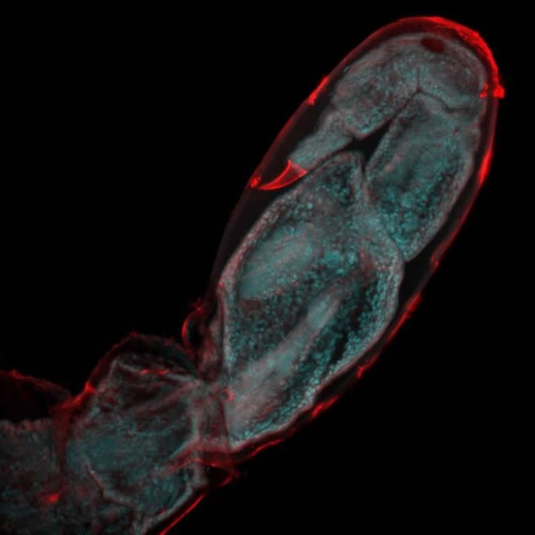
In a study to be published in the journal eLife, the team, from the Institute of Functional Genomics in Lyon (Institut de Génomique Fonctionnelle de Lyon, IGFL), France, carried out continuous live imaging of a regenerating leg in the crustacean Parhyale hawaiensis, a close relative of the common sand hopper.
Parhyale hawaiensis was introduced in 2014 as a model for regenerative studies in a paper co-authored by Michalis Averof, Director of Research at IGFL and senior author of the current study.
“Parhyale hawaiensis is well suited for imaging limb regrowth,” Averof says. “The animals have relatively rapid limb regeneration, requiring as little as one week for young adults to fully regrow their legs. Also, their tiny limbs enable us to image the regeneration process in unprecedented cell-by-cell detail through their entire thickness.”
To record the regrowth of the Parhyale hawaiensis leg, the team used genetically engineered tools to label the limb’s epidermal cells using fluorescent proteins. They then used microscopy to take continuous live recordings over the first days of regrowth, with the fluorescence in the cells allowing them to capture their individual activity.
“Using this method, we identified a specific sequence of events and cell behaviours that unfold during limb regrowth,” says first author Frederike Alwes.
“These include wound closure, followed by a quiet period when the epidermal cells migrate slowly towards the site of the wound, which then leads to extensive cell division and movement as the new leg starts to develop its shape. We were surprised to see that there was a sharp transition between the last two stages, which suggests the cells were coordinated by a common signal.”
The study also showed for the first time that there are no specialised stem cells for regenerating the epidermis in new limbs. Instead, most of the leg stump’s epidermal cells divide and re-arrange, contributing to new segments in the leg.
Alwes explains: “Traditionally, insight into cell behaviour during limb regrowth has been gathered by imaging fixed samples and attempting to fill in the missing pieces, due to the difficulties in tracking cells during regeneration in active adult animals. With the ability to track the movements and behaviour of single cells individually through time, we now have the means to understand the cellular dynamics of the regeneration process, which could not have been reconstructed from fixed material.
“While our paper focuses mostly on the behaviour of epidermal cells, we now plan to extend this work to include all the different cell types that are involved in limb regrowth. The ultimate aim of our research is to explore how some animals can respond to a severe injury by regenerating an entire body part that was lost.”
Funding: Funding provided by Agence Nationale de la Recherche.
Source: Emily Packer – eLife
Image Source: NeuroscienceNews.com image is credited to Frederike Alwes.
Video Source: Video is credited to Carmen Ferguson.
Original Research: Full open access research for “Live imaging reveals the progenitors and cell dynamics of limb regeneration” by Frederike Alwes, Camille Enjolras, and Michalis Averof in eLife. Published online October 25 2016 doi:10.7554/eLife.19766
[cbtabs][cbtab title=”MLA”]eLife “First Time Lapse Footage of Cell Activity During Limb Regeneration.” NeuroscienceNews. NeuroscienceNews, 25 October 2016.
<https://neurosciencenews.com/limb-regeneration-time-lapse-5351/>.[/cbtab][cbtab title=”APA”]eLife (2016, October 25). First Time Lapse Footage of Cell Activity During Limb Regeneration. NeuroscienceNew. Retrieved October 25, 2016 from https://neurosciencenews.com/limb-regeneration-time-lapse-5351/[/cbtab][cbtab title=”Chicago”]eLife “First Time Lapse Footage of Cell Activity During Limb Regeneration.” https://neurosciencenews.com/limb-regeneration-time-lapse-5351/ (accessed October 25, 2016).[/cbtab][/cbtabs]
Abstract
Live imaging reveals the progenitors and cell dynamics of limb regeneration
Regeneration is a complex and dynamic process, mobilizing diverse cell types and remodelling tissues over long time periods. Tracking cell fate and behaviour during regeneration in active adult animals is especially challenging. Here, we establish continuous live imaging of leg regeneration at single-cell resolution in the crustacean Parhyale hawaiensis. By live recordings encompassing the first 4-5 days after amputation, we capture the cellular events that contribute to wound closure and morphogenesis of regenerating legs with unprecedented resolution and temporal detail. Using these recordings we are able to track cell lineages, to generate fate maps of the blastema and to identify the progenitors of regenerated epidermis. We find that there are no specialized stem cells for the epidermis. Most epidermal cells in the distal part of the leg stump proliferate, acquire new positional values and contribute to new segments in the regenerating leg.
“Live imaging reveals the progenitors and cell dynamics of limb regeneration” by Frederike Alwes, Camille Enjolras, and Michalis Averof in eLife. Published online October 25 2016 doi:10.7554/eLife.19766