

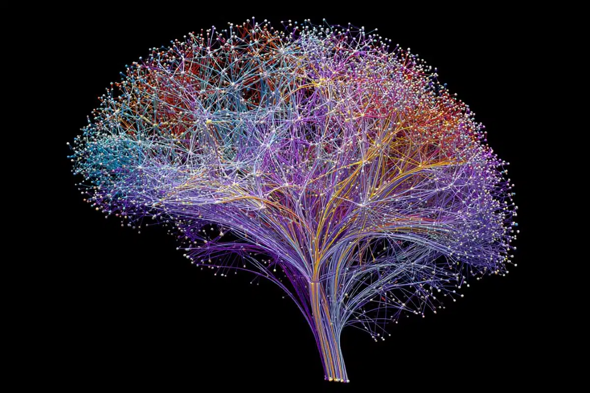
Summary: Researchers engineered one of the largest reference models ever constructed for the human brain. The study compiled diffusion MRI scans from 54,583 individuals across 19 international datasets to establish definitive, lifelong “growth and decline charts” for the brain’s white matter.
By tracking how water molecules travel along neural wiring across the lifespan, this publicly available tool enables clinicians to map person-specific structural deviations linked to aging, Alzheimer’s disease, and schizophrenia risk.
Key Facts
- The White Matter Growth Metric: Much like standard pediatric growth charts track a child’s height and weight milestones, this framework provides a normative reference standard for the brain’s white matter microstructure. It maps out exactly how the vast network of neural wiring matures and declines over time, offering a new method to detect subtle structural abnormalities.
- The Diffusion MRI Lens: To map this microstructural terrain on a global scale, the team utilized diffusion MRI, a specialized imaging technique that monitors the microscopic movement of water through living brain tissue. Because this fluid movement is physically steered by nerve fibers and the density of their protective myelin coating, the scans catch structural shifts entirely invisible on standard clinical brain scans.
- Validating the “Last In, First Out” Axiom: The global datasets yielded a massive structural insight, validating a long-standing theory of brain aging known as “last in, first out”. The team observed that the specific white matter pathways that take the longest to fully mature in childhood and adolescence are the exact same neural highways that exhibit the fastest structural decline as the brain encounters old age.
- Person-Specific Diagnostic Deviations: To test the practical clinical utility of the charts, the team applied their normative model to datasets from individuals diagnosed with mild cognitive impairment, dementia, and 22q11.2 deletion syndrome (a genetic variant indicating high schizophrenia risk). In every single case, the model flagged explicit structural deviations from age-expected norms. Crucially, these deviations were not identical among individuals with the same diagnosis, highlighting the necessity of an individualized approach to brain health.
- The Clinical Trial Dashboard: Spearheaded by senior author Dr. Paul M. Thompson and first author Dr. Julio E. Villalón-Reina, the charts allow scientists to cross-evaluate an individual’s neural pathways relative to other people of the exact same age, sex, and demographic cohort. This delivers a baseline standard to evaluate treatment trials, tracking whether a therapeutic intervention successfully pulls a patient’s white matter metrics back toward healthy ranges or slows down degeneration over time.
- A Common Framework for 30+ Disorders: Funded by the National Institutes of Health (NIH) and international partners, the publicly available reference tool is currently being scaled to rigorously compare more than 30 separate neurological, psychiatric, and neurodevelopmental conditions under a single, unified analytical framework.
Source: USC
Researchers at the USC Mark and Mary Stevens Neuroimaging and Informatics Institute (Stevens INI) at the Keck School of Medicine of USC have created one of the largest reference models ever developed for the human brain, using diffusion MRI scans from more than 54,000 people to chart how the brain’s communication pathways develop, mature, and decline across the lifespan.
Published in Nature Communications, the study provides the equivalent of “growth charts” for the brain’s white matter – the vast network of neural wiring that allows brain regions to communicate. The new tool offers researchers a new way to detect subtle patterns linked to aging, Alzheimer’s disease, schizophrenia risk, and other neurological and psychiatric conditions.
“Just as pediatric growth charts help clinicians determine whether a child’s height or weight is developing as expected, these brain charts provide a reference for how the brain’s neural pathways typically change over the lifespan,” said Julio E. Villalón-Reina, MD, PhD, a postdoctoral researcher at the Stevens INI and the study’s first author. “That gives us a powerful new way to identify when an individual’s brain wiring falls outside the expected range.”
White matter is essential for efficient communication throughout the brain. To study it, the team used diffusion MRI, an imaging method that tracks how water moves through brain tissue. Because water movement is shaped by microscopic features such as nerve fibers and myelin, the protective coating around them, diffusion MRI can reveal subtle changes in tissue organization not visible on standard brain scans.
After compiling diffusion MRI data from 54,583 individuals across 19 international datasets, the researchers built statistical “growth and decline charts” for the brain’s neural pathways, the network of nerve fibers that connects different regions of the brain and allows them to communicate.
The researchers focused on four widely used measures of white matter microstructure across 21 major brain regions. By modeling how these measures vary by age and sex, they generated lifespan curves and percentile ranges that show what is typical at different stages of life. The results revealed that white matter follows distinct developmental and aging trajectories, with some measures reaching peak maturity in early adulthood and others later in midlife.
“Brain development and brain aging are not uniform processes,” Villalón-Reina said. “The brain’s neural pathways mature on distinct timelines, and some are more vulnerable to decline than others. Our model reveals this structure by merging data on a truly global scale.”
The team also discovered evidence for a longstanding theory of brain aging, sometimes described as “last in, first out.” According to this theory, brain pathways that develop last in childhood and adolescence tend to be more susceptible to decline in older age. The researchers observed that white matter regions that mature later did indeed decline faster in old age, offering new insight linking brain development and aging.
To demonstrate the model’s practical value, the researchers applied it to clinical datasets from people with mild cognitive impairment, dementia, and 22q11.2 deletion syndrome, a genetic condition that increases risk of schizophrenia. In each case, the model identified alterations in the brain’s circuitry that deviated from age-expected norms. Importantly, these deviations were not identical across individuals with the same diagnosis, highlighting the value of a person-specific approach.
“This monumental study took seven years to complete,” said Paul M. Thompson, PhD, associate director of the Stevens INI and senior author of the study.
“The vast scale of the data and the fine scale of the brain features assessed means we can now evaluate your neural pathways relative to other people of the same age, sex, and demographics. We can see how your brain differs from what we would expect for a person of your age and sex, giving us a tool to use in clinical trials of treatments for dozens of brain diseases.”
When applied to people with dementia and mild cognitive impairment, the model detected atypical white matter patterns in brain regions involved in memory and interregional communication. In people with 22q11.2 deletion syndrome, it identified deviations in multiple key neural pathways, helping researchers discover which brain systems develop differently.
The reference charts may also help researchers evaluate treatments by tracking whether a person’s white matter measures move closer to the expected range, or whether a treatment slows the shift away from healthy patterns over time. The charts will now be used to compare more than 30 brain diseases and conditions, offering a common framework for studying how different disorders emerge, progress, and respond to intervention.
The models are also a publicly available resource that can be extended as additional brain imaging data become available. The methods are now being used to study neurological, psychiatric, and neurodevelopmental disorders by providing a common reference standard for white matter microstructure across the lifespan.
“This study demonstrates the power of large-scale, international data sharing to create tools the entire research community can use,” said Arthur W. Toga, PhD, director of the Stevens INI and Provost Professor at USC. “By establishing a lifespan framework for the brain’s communication pathways, this work opens new opportunities to detect subtle disease-related changes, compare conditions more rigorously, and move toward a more individualized understanding of brain health.”
About the study
The study, “Lifespan normative modeling of brain microstructure,” was published in Nature Communications. In addition to Villalón-Reina and Thompson, the study’s authors include Alyssa H. Zhu, Leila Nabulsi, Sophia I. Thomopoulos, Clara A. Moreau, Yixue Feng, Tamoghna Chattopadhyay, Sebastian Benavidez, Leila Kushan, John P. John, Himanshu Joshi, Iyad Ba Gari, Katherine E. Lawrence, Talia M. Nir, Neda Jahanshad, Carrie E. Bearden, Seyed Mostafa Kia, Andre F. Marquand and the Alzheimer’s Disease Neuroimaging Initiative.
Funding: This work was supported by grants from the National Institutes of Health, including the National Institute on Aging, the Fogarty International Center and the National Institute of Mental Health, as well as support from the Alzheimer’s Association, the European Research Council and the Wellcome Trust, and the Popovich Chair in Neurodegenerative Diseases.
Key Questions Answered:
A: By giving doctors a definitive percent-range reference to judge if a person’s brain structure matches their chronological age. Just like pediatricians use charts to see if a baby’s height falls in the 50th or 90th percentile, this USC model allows neuroscientists to look at four key measures of white matter across 21 major brain regions. It plots out an expected baseline, revealing instantly if an individual’s neural wiring is aging normally or falling off the expected curve.
A: It means that the last parts of our brain to grow up are the very first parts to break down. The study confirmed that the complex neural pathways that finish developing latest in childhood and adolescence are biologically the most vulnerable systems. As we step into old age, these late-maturing white matter tracts undergo the fastest structural decline, linking the timelines of development and aging.
A: Because neurological diseases do not print identical structural copies across different human bodies. When the researchers applied their model to clinical datasets, they discovered that even when people shared the same condition, like dementia or schizophrenia risk, their specific white matter deviations from age-expected norms were unique. This proves that brain health requires a personalized, person-specific framework to ensure precision treatments.
Editorial Notes:
- This article was edited by a Neuroscience News editor.
- Journal paper reviewed in full.
- Additional context added by our staff.
About this neuroscience and brain mapping research news
Author: Laura LeBlanc
Source: USC
Contact: Laura LeBlanc – USC
Image: The image is credited to Neuroscience News
Original Research: Open access.
“Lifespan normative modeling of brain microstructure” by Julio E. Villalón-Reina, Alyssa H. Zhu, Leila Nabulsi, Sophia I. Thomopoulos, Clara A. Moreau, Yixue Feng, Tamoghna Chattopadhyay, Sebastian M. Benavidez, Leila Kushan, John P. John, Himanshu Joshi, Iyad Ba Gari, Katherine E. Lawrence, Talia M. Nir, Neda Jahanshad, Carrie E. Bearden, Seyed Mostafa Kia, Andre F. Marquand, the Alzheimer’s Disease Neuroimaging Initiative & Paul M. Thompson. Nature Communications
DOI:10.1038/s41467-026-72875-x
Abstract
Lifespan normative modeling of brain microstructure
Normative models of brain metrics based on large populations could be extremely valuable for detecting brain abnormalities in patients with a variety of disorders, including degenerative, psychiatric and neurodevelopmental conditions, but no such models exist for the brain’s white matter (WM) microstructure.
Here we present a large-scale normative model of brain WM microstructure – based on 19 international diffusion MRI datasets covering almost the entire lifespan (totaling N = 54,583 individuals; age: 4–91 years). We extracted regional diffusion tensor imaging (DTI) metrics using a standardized analysis and quality control protocol and used hierarchical Bayesian regression (HBR) to model the statistical distribution of derived WM metrics as a function of age and sex. We extracted the average lifespan trajectories and corresponding centile curves for each WM region.
We illustrate the utility of the method by applying it to detect and visualize profiles of WM microstructural deviations in a variety of contexts: in mild cognitive impairment, Alzheimer’s disease, and 22q11.2 deletion syndrome – a neurogenetic condition that markedly increases risk for schizophrenia.
The resulting large-scale model provides a common reference to identify disease effects on the brain’s microstructure in individuals or groups, and to compare disorders, and discover factors affecting WM abnormalities.
The derived normative models are a valuable resource publicly available to the community, adaptable and extendable to future datasets as the available data expands.


