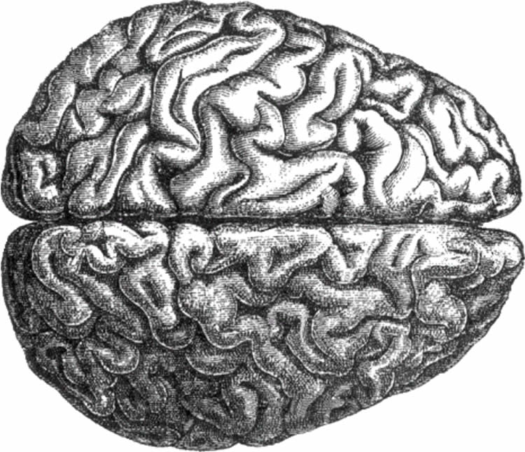
Neuroimaging studies suggest that frontolimbic regions of the brain, structures that regulate emotions, play an important role in the biology of aggressive behavior.
A new article published in the inaugural issue of the journal Biological Psychiatry: Cognitive Neuroscience and Neuroimaging reports that individuals with intermittent explosive disorder (IED) have significantly lower gray matter volume in these frontolimbic brain structures. In other words, these people have smaller “emotional brains.”
“Intermittent explosive disorder is defined in DSM-5 as recurrent, problematic, impulsive aggression,” explained Dr. Emil Coccaro, the article’s lead author. “While more common than bipolar disorder and schizophrenia combined, many in the scientific and lay communities believe that impulsive aggression is simply ‘bad behavior’ that requires an ‘attitude adjustment.’ However, our data confirm that IED, as defined by DSM-5, is a brain disorder and not simply a disorder of ‘personality.'” Dr. Coccaro is the E.C. Manning Professor and Chair of Psychiatry and Behavioral Neuroscience at the University of Chicago.

Dr. Coccaro and his colleagues also report a significant inverse correlation between measures of aggression and frontolimbic gray matter volume.
The investigators collected high-resolution magnetic resonance imaging (MRI) scans in 168 subjects, including 57 subjects with IED, 53 healthy control subjects, and 58 psychiatric control subjects. The team found a direct correlation between history of actual aggressive behavior and the magnitude of reduction in gray matter volume, linking both in a dimensional relationship.
“Across all subjects, reduced volume in frontolimbic brain structures was associated with increased aggressiveness,” commented Dr. Cameron Carter, Professor of Psychiatry and Behavioral Sciences at University of California, Davis and Editor of Biological Psychiatry: Cognitive Neuroscience and Neuroimaging. “These important findings suggest that disrupted development of the brain’s emotion-regulating circuitry may underlie an individual’s propensity for rage and aggression.”
Source: Rhiannon Bugno – Elsevier
Image Source: The image is in the public domain
Original Research: Abstract for “Frontolimbic Morphometric Abnormalities in Intermittent Explosive Disorder and Aggression” by Emil F. Coccaro, Daniel A. Fitzgerald, Royce Lee, Michael McCloskey, and K. Luan Phan in Biological Psychiatry: Cognitive Neuroscience and Neuroimaging. Published online January 2016 doi:10.1016/j.bpsc.2015.09.006
Abstract
Frontolimbic Morphometric Abnormalities in Intermittent Explosive Disorder and Aggression
Background
Converging evidence from neuroimaging studies suggests that impulsive aggression, the core behavior in the DSM-5 diagnosis intermittent explosive disorder (IED), is regulated by frontolimbic brain structures, particularly orbitofrontal cortex, ventral medial prefrontal cortex, anterior cingulate cortex, amygdala, insula, and uncus. Despite this evidence, no brain volumetric studies of IED have been reported as yet. This study was conducted to test the hypothesis that gray matter volume in frontolimbic brain structures of subjects with IED is lower than in healthy subjects and subjects with other psychiatric conditions.
Methods
High-resolution magnetic resonance imaging scans using a three-dimensional magnetization-prepared rapid acquisition gradient-echo sequence were performed in 168 subjects (n = 53 healthy control subjects, n = 58 psychiatric controls, n = 57 subjects with IED). Imaging data were analyzed by voxel-based morphometry methods employing Statistical Parametric Mapping (SPM8) software.
Results
Gray matter volume was found to be significantly lower in subjects with IED compared with healthy control subjects and psychiatric controls in orbitofrontal cortex, ventral medial prefrontal cortex, anterior cingulate cortex, amygdala, insula, and uncus. These differences were not due to various confounding factors or to comorbidity with other disorders previously reported to have reduced gray matter volume. Gray matter volume in these areas was significantly and inversely correlated with measures of aggression.
Conclusions
Reductions in the gray matter volume of frontolimbic structures may be a neuronal characteristic of impulsively aggressive individuals with DSM-5 IED. These data suggest an anatomic correlate accounting for functional deficits in social-emotional information processing in these individuals.
“Frontolimbic Morphometric Abnormalities in Intermittent Explosive Disorder and Aggression” by Emil F. Coccaro, Daniel A. Fitzgerald, Royce Lee, Michael McCloskey, and K. Luan Phan in Biological Psychiatry: Cognitive Neuroscience and Neuroimaging. Published online January 2016 doi:10.1016/j.bpsc.2015.09.006


