Summary: Neuroimaging reveals areas of the brain associated with visual and auditory processing are more active when anxiety slowly increases during horror movies. After a shocking scene, brain areas associated with emotional processing, threat evaluation, and decision making increase in activity.
Source: University of Turku
Finnish research team maps neural activity in response to watching horror movies. A study conducted by the University of Turku shows the top horror movies of the past 100 years, and how they manipulate brain activity.
Humans are fascinated by what scares us, be it sky-diving, roller-coasters, or true-crime documentaries – provided these threats are kept at a safe distance. Horror movies are no different.
Whilst all movies have our heroes face some kind of threat to their safety or happiness, horror movies up the ante by having some kind of superhuman or supernatural threat that cannot be reasoned with or fought easily.
The research team at the University of Turku, Finland, studied why we are drawn to such things as entertainment? The researchers first established the 100 best and scariest horror movies of the past century, and how they made people feel.
Unseen Threats Are Most Scary
Firstly, 72% of people report watching at last one horror movie every 6 months, and the reasons for doing so, besides the feelings of fear and anxiety, was primarily that of excitement. Watching horror movies was also an excuse to socialise, with many people preferring to watch horror movies with others than on their own.
People found horror that was psychological in nature and based on real events the scariest, and were far more scared by things that were unseen or implied rather than what they could actually see.
“This latter distinction reflects two types of fear that people experience. The creeping foreboding dread that occurs when one feels that something isn’t quite right, and the instinctive response we have to the sudden appearance of a monster that make us jump out of our skin, says principal investigator,” Professor Lauri Nummenmaa from Turku PET Centre.
MRI Reveals How Brain Reacts to Different Forms of Fear
Researchers wanted to know how the brain copes with fear in response to this complicated and ever changing environment. The group had people watch a horror movie whilst measuring neural activity in a magnetic resonance imaging scanner.
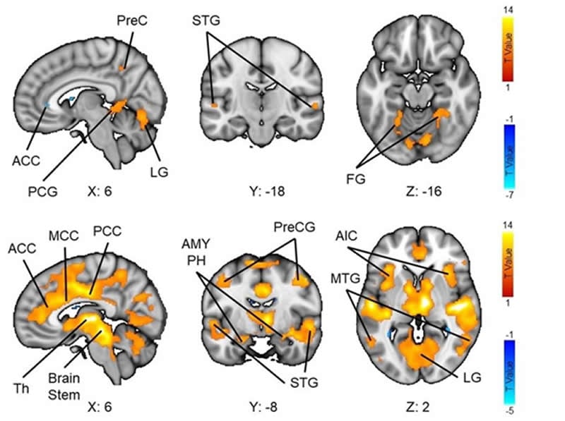
During those times when anxiety is slowly increasing, regions of the brain involved in visual and auditory perception become more active, as the need to attend for cues of threat in the environment become more important. After a sudden shock, brain activity is more evident in regions involved in emotion processing, threat evaluation, and decision making, enabling a rapid response.
However, these regions are in continuous talk-back with sensory regions throughout the movie, as if the sensory regions were preparing response networks as a scary event was becoming increasingly likely.
“Therefore, our brains are continuously anticipating and preparing us for action in response to threat, and horror movies exploit this expertly to enhance our excitement,” explains Researcher Matthew Hudson.
The horror movie survey is still active. You may participate here.
Funding: This research was funded by NIA grant R01AG053155.
Source:
University of Turku
Media Contacts:
Lauri Nummenmaa – University of Turku
Image Source:
The image is credited to Lauri Nummenmaa.
Original Research: Open access
“Dissociable neural systems for unconditioned acute and sustained fear”. Lauri Nummenmaa, et al.
Neurolmage doi:10.1016/j.neuroimage.2020.116522.
Abstract
Dissociable neural systems for unconditioned acute and sustained fear
Fear protects organisms by increasing vigilance and preparedness, and by coordinating survival responses during life-threatening encounters. The fear circuit must thus operate on multiple timescales ranging from preparatory sustained alertness to acute fight-or-flight responses. Here we studied the brain basis of sustained and acute fear using naturalistic functional magnetic resonance imaging (fMRI) enabling analysis of different time-scales of fear responses. Subjects (N = 37) watched feature-length horror movies while their hemodynamic brain activity was measured with fMRI. Time-variable intersubject correlation (ISC) was used to quantify the reliability of brain activity across participants, and seed-based phase synchronization was used for characterizing dynamic connectivity. Subjective ratings of fear were used to assess how synchronization and functional connectivity varied with emotional intensity. These data suggest that acute and sustained fear are supported by distinct neural pathways, with sustained fear amplifying mainly sensory responses, and acute fear increasing activity in brainstem, thalamus, amygdala and cingulate cortices. Sustained fear increased ISC in regions associated with acute fear, and also amplified functional connectivity within this network. The results were replicated in an independent experiment with a different subject sample and stimulus movie. The functional interplay between cortical networks involved in sustained anticipation of, and acute response to, threat involves a complex and dynamic interaction that depends on the proximity of threat, and the need to employ threat appraisals and vigilance for decision making and response selection.