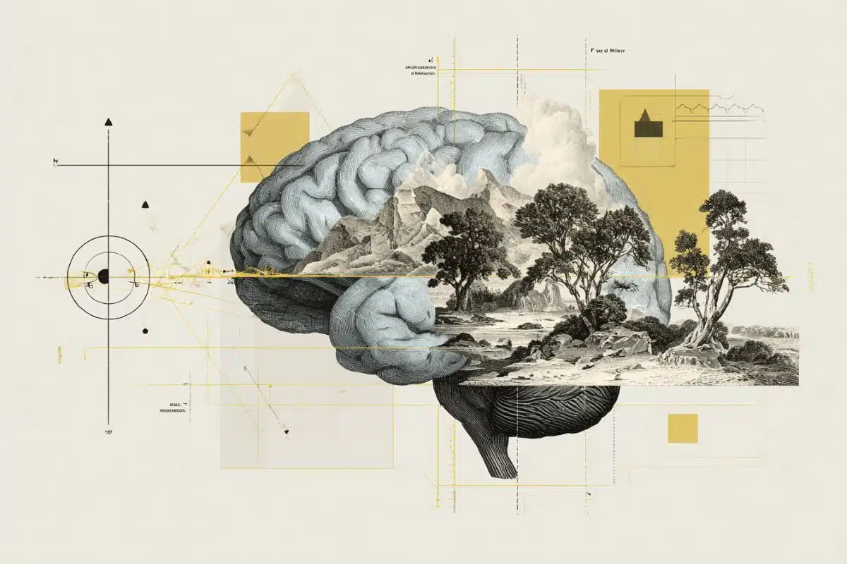
Summary: The hippocampus is often called the “GPS of the brain,” but a new study reveals it is much more than a simple map. The research shows that the hippocampus acts like a dynamic dial that shifts its activity based on whether our expectations match reality. When everything is as expected, brain activity flows smoothly across the structure.
However, when something changes, the hippocampus physically partitions itself: the front (anterior) handles “What” (conceptual changes), while the back (posterior) handles “Where” (spatial changes). This discovery proves that the brain uses a flexible architecture to reconcile meaning and location in real-time.
Key Facts
- The expectation Dial: When a sequence of events matches a person’s memory, hippocampal activity moves in a continuous, smooth wave from front to back.
- The “What” Region (Anterior): If a person expects to see one object (a dog) but sees another (a cat), the front of the hippocampus lights up. This area is connected to systems for abstract and conceptual processing.
- The “Where” Region (Posterior): If an object appears in the wrong location, the back of the hippocampus takes over. This area is linked to visual and spatial processing networks.
- The Reconciliation Center: When both the object and the location change, activity centers in the middle of the hippocampus, suggesting this central zone merges spatial and conceptual data.
- Beyond the GPS: While the 2014 Nobel Prize highlighted “place cells” for navigation, this study suggests the hippocampus is equally focused on semantic meaning, the “why” and “what” of our environment.
Source: University of Chicago
The hippocampus is a crucial part of the brain that plays a role in memory and learning, especially in remembering directions and locations.
New research from the University of Chicago shows how this small, curved structure reorganizes its activity depending on whether a situation matches people’s memories and expectations.
In a new study published in the Proceedings of the National Academy of Sciences, researchers used functional MRI scanning to track the brain activity of study participants as they watched images of a sequence of objects at different locations.
When the images matched what the participants expected, activity in the hippocampus shifted smoothly from the front to the back, like a continuous dial. When the images differed from the patterns the subjects had learned, however, the researchers saw that hippocampal activity split into specialized regions:
- If the “what” of the image changed—for example, the participant expected to see a picture of a dog but instead saw a cat—the activity took place in the anterior, or front, part of the hippocampus.
- If the “where” of the image changed—the picture of the dog was on the left side instead of the right—activity centered in the posterior, or back, part of the hippocampus.
“Real memories involve more than just objects or locations. They are bound to concepts and meaning. How the hippocampus handles both space and meaning at the same time has been one of the central unsolved questions in memory neuroscience,” said James Kragel, PhD, a Research Assistant Professor in the Department of Neurology at UChicago and senior author of the study.
“This resolves a long-standing debate about hippocampal organization and suggests that flexibility, not fixed architecture, is a core principle of how the brain organizes memory, spanning both spatial and semantic information.”
The GPS of the brain
Sometimes called “the GPS of the brain,” the hippocampus is most well-known for its role in remembering places and locations. In 2014, the Nobel Prize in Medicine was awarded for the discovery of so-called place cells and grid cells in the brain that track location and map out space.
A lot of research on the hippocampus to date has focused on these spatial aspects; other MRI studies in rodents and humans suggest that the hippocampus processes big-picture information in the anterior region, and more precise details in the posterior.
But just as important as spatial information is what’s in those locations. Our experience of a room is distinctly different if all the furniture is in the same place, but the couch in the corner is now bright red instead of blue, as we remembered. Kragel and his teammates wanted to go further to understand the overlap of how spatial and conceptual information are represented at the same time.
They recruited 28 participants who learned sequences of five images placed in different locations on a circular array. After they memorized the sequences, they got into an MRI machine and watched a replay of those same images—with some differences. The researchers varied the sequences, sometimes swapping an image in its expected location, moving an image to a different location, or both.
Depending on how big the difference was from the expected images, the researchers saw different activity in the hippocampus. Seeing an image of a different object triggered more activity in the anterior region, while differences in location sparked more activity in the posterior region. Changes in both the object and its location generated activity in the central region, suggesting that it plays a role in reconciling both types of information.
Sorting and responding
Different regions of the hippocampus are connected to different networks of the brain for higher-level processing. The anterior region is connected to systems involved in abstract or conceptual processing, and the posterior region is connected to systems for visual and spatial processing.
This suggests that the patterns of activity the researchers saw in this study are a way for the hippocampus to sort out discrepancies and pass them along to more specialized parts of the brain for further processing.
“We need to encode and retrieve memories pretty quickly all the time, and we need to be able to switch between processing different types of information,” Kragel said.
“So, this type of organization where the hippocampus receives different kinds of inputs allows it to rapidly detect when information differs from our expectations and retrieve relevant memories to guide behavior.”
Funding: The study, “Spatial and semantic memory reorganize a hippocampal long-axis gradient,” was supported by the National Institutes of Health.
Additional authors include Anikka G. Jordan and Joel L. Voss, both from UChicago.
Key Questions Answered:
A: It’s about processing speed and specialized networks. The front of your brain is better at “big picture” concepts and meaning, while the back is optimized for 3D space and visual precision. By splitting these tasks, the hippocampus can quickly tell the rest of your brain exactly what part of your memory needs to be updated without re-scanning the entire scene.
A: If the hippocampus couldn’t shift between these regions, you might experience “reality discrimination” issues. You might recognize a friend (the “what”) but feel a deep sense of unease because your brain can’t reconcile that they are in your kitchen instead of their office (the “where”). This flexibility is key to feeling “grounded” in your surroundings.
A: Your brain is a prediction machine. The smooth “front-to-back” flow of activity seen in the study is the sound of a brain whose predictions are coming true. When that flow breaks, it’s a “red alert” signal that tells your brain to stop coasting on autopilot and start encoding a new, unexpected memory.
Editorial Notes:
- This article was edited by a Neuroscience News editor.
- Journal paper reviewed in full.
- Additional context added by our staff.
About this neuroscience research news
Author: Cassandra Belek
Source: University of Chicago
Contact: Cassandra Belek – University of Chicago
Image: The image is credited to Neuroscience News
Original Research: Closed access.
“Spatial and semantic memory reorganize a hippocampal long-axis gradient” by Anikka G. Jordan, Joel L. Voss, and James E. Kragel. PNAS
DOI:10.1073/pnas.2525724123
Abstract
Spatial and semantic memory reorganize a hippocampal long-axis gradient
The hippocampus supports episodic memory by binding spatial and semantic information, yet how this information is simultaneously organized along its long axis remains debated.
Gradient accounts propose a continuous shift in representational scale, from coarse coding in anterior to fine coding in posterior regions, whereas modular accounts posit discrete subregions specialized for distinct functions.
Using high-resolution fMRI together with eye tracking as a readout of spatial and semantic memory during sequence learning, we directly tested these competing models.
During predictable sequences, hippocampal activity continuously varied along the long axis. In contrast, modular organization emerged when sequences mismatched memory.
Subregions in the anterior and posterior hippocampus were sensitive to semantic and spatial mismatches, respectively. Notably, the intermediate hippocampus was specifically sensitive to concurrent mismatches in both dimensions, but not to mismatches in either dimension alone.
These content-sensitive subregions were embedded within distinct cortical networks that reorganized according to memory demands.
Together, our findings reveal a dynamic hippocampal architecture that flexibly combines gradient and modular principles to simultaneously represent the spatial and semantic content that defines episodic memory.